

Irradiation induces bone injury by damaging bone marrow microenvironment for stem cells
- PMID: 21220327
- PMCID: PMC3029740
- DOI: 10.1073/pnas.1015350108
Irradiation induces bone injury by damaging bone marrow microenvironment for stem cells
Erratum in
- Proc Natl Acad Sci U S A. 2011 Apr 5;108(14):5921
Abstract
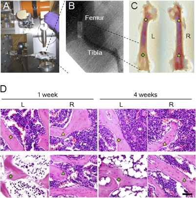
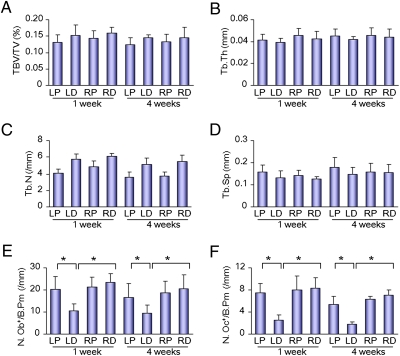
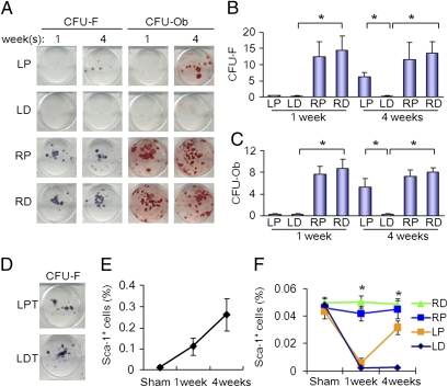
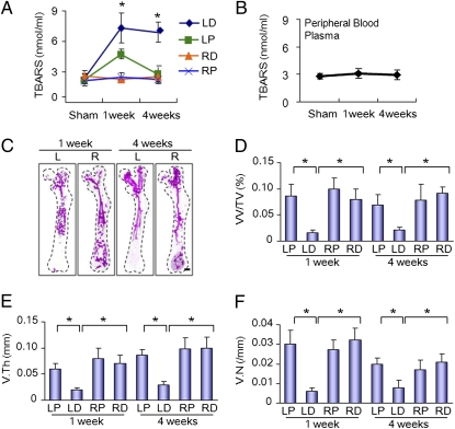
Radiation therapy can result in bone injury with the development of fractures and often can lead to delayed and nonunion of bone. There is no prevention or treatment for irradiation-induced bone injury. We irradiated the distal half of the mouse left femur to study the mechanism of irradiation-induced bone injury and found that no mesenchymal stem cells (MSCs) were detected in irradiated distal femora or nonirradiated proximal femora. The MSCs in the circulation doubled at 1 week and increased fourfold after 4 wk of irradiation. The number of MSCs in the proximal femur quickly recovered, but no recovery was observed in the distal femur. The levels of free radicals were increased threefold at 1 wk and remained at this high level for 4 wk in distal femora, whereas the levels were increased at 1 wk and returned to the basal level at 4 wk in nonirradiated proximal femur. Free radicals diffuse ipsilaterally to the proximal femur through bone medullary canal. The blood vessels in the distal femora were destroyed in angiographic images, but not in the proximal femora. The osteoclasts and osteoblasts were decreased in the distal femora after irradiation, but no changes were observed in the proximal femora. The total bone volumes were not affected in proximal and distal femora. Our data indicate that irradiation produces free radicals that adversely affect the survival of MSCs in both distal and proximal femora. Irradiation injury to the vasculatures and the microenvironment affect the niches for stem cells during the recovery period.
Conflict of interest statement
The authors declare no conflict of interest.
Figures
References
-
- Costantino PD, Friedman CD, Steinberg MJ. Irradiated bone and its management. Otolaryngol Clin North Am. 1995;28:1021–1038. - PubMed
-
- Jegoux F, Malard O, Goyenvalle E, Aguado E, Daculsi G. Radiation effects on bone healing and reconstruction: interpretation of the literature. Oral Surg Oral Med Oral Pathol Oral Radiol Endod. 2010;109:173–184. - PubMed
-
- Baxter NN, Habermann EB, Tepper JE, Durham SB, Virnig BA. Risk of pelvic fractures in older women following pelvic irradiation. JAMA. 2005;294:2587–2593. - PubMed
-
- Gerson SL. Mesenchymal stem cells: No longer second class marrow citizens. Nat Med. 1999;5:262–264. - PubMed
-
- Prockop DJ. Marrow stromal cells as stem cells for nonhematopoietic tissues. Science. 1997;276:71–74. - PubMed
MeSH terms
Substances
LinkOut - more resources
Full Text Sources
Other Literature Sources
