. 2011 Jan 25;108(4):1639-44.
doi: 10.1073/pnas.1013888108. Epub 2011 Jan 10.

Translocation of surface-localized effectors in type III secretion

Affiliations

Translocation of surface-localized effectors in type III secretion

Karen Akopyan et al. Proc Natl Acad Sci U S A. .

Abstract

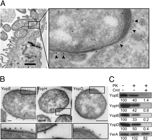
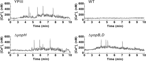
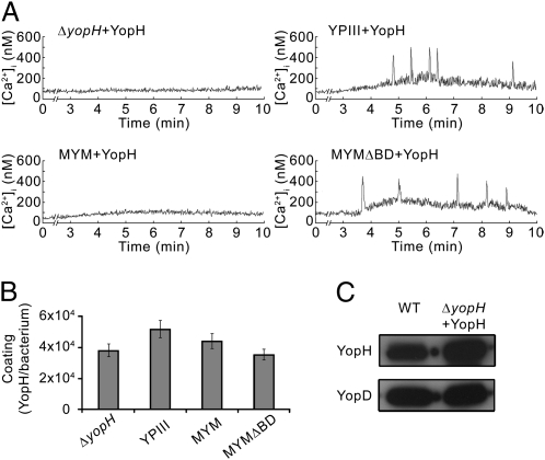
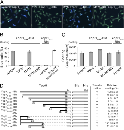
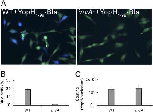
Pathogenic Yersinia species suppress the host immune response by using a plasmid-encoded type III secretion system (T3SS) to translocate virulence proteins into the cytosol of the target cells. T3SS-dependent protein translocation is believed to occur in one step from the bacterial cytosol to the target-cell cytoplasm through a conduit created by the T3SS upon target cell contact. Here, we report that T3SS substrates on the surface of Yersinia pseudotuberculosis are translocated into target cells. Upon host cell contact, purified YopH coated on Y. pseudotuberculosis was specifically and rapidly translocated across the target-cell membrane, which led to a physiological response in the infected cell. In addition, translocation of externally added YopH required a functional T3SS and a specific translocation domain in the effector protein. Efficient, T3SS-dependent translocation of purified YopH added in vitro was also observed when using coated Salmonella typhimurium strains, which implies that T3SS-mediated translocation of extracellular effector proteins is conserved among T3SS-dependent pathogens. Our results demonstrate that polarized T3SS-dependent translocation of proteins can be achieved through an intermediate extracellular step that can be reconstituted in vitro. These results indicate that translocation can occur by a different mechanism from the assumed single-step conduit model.


Conflict of interest statement

The authors declare no conflict of interest.

Figures

Fig. 1.
Fig. 1.
YopE is evenly distributed in the bacterium-target cell interface during infection of HeLa cells and Yops are found on the surface of Y. pseudotuberculosis before target cell contact. (A) Immunoelectron micrograph showing localization of YopE in Y. pseudotuberculosis during infection of a HeLa cell. Arrowheads indicate YopE detected on the surface of or within the plasma membrane of the target cell. (B) Immunoelectron micrographs showing surface localization of YopE, YopH, and YopD on Y. pseudotuberculosis before target-cell contact. (Scale bars, 100 nm for YopE and YopH; 200 nm for YopD.) (C) Western blot analysis of YopE, YopH, YopB, YopD, and the cytoplasmic T3SS chaperone YerA (SycE) in Y. pseudotuberculosis treated with chloramphenicol (Cml) or proteinase K (PK). Relative signal intensities are indicated below each panel.
Fig. 2.
Fig. 2.
YopH-mediated inhibition of the immediate-early Ca2+ response in human neutrophils. Real-time monitoring of intracellular Ca2+ in neutrophils infected with chloramphenicol-treated Y. pseudotuberculosis strains: plasmid-cured mutant (YPIII), wild-type bacteria (WT), a yopH deletion mutant (ΔyopH), and a double yopByopD-deletion mutant (ΔyopB,D). The mean numbers (± SEM) of Ca2+ peaks (300–600 nM) generated within 10 min of infection in nine independent experiments were as follows: YPIII, 3.41 ± 0.62; wild type, 0.22 ± 0.10; ΔyopH, 3.39 ± 0.53; ΔyopB+D, 3.28 ± 0.81.
Fig. 3.
Fig. 3.
Y. pseudotuberculosis yopH mutant strains coated with purified YopH block the immediate-early Ca2+ response in human neutrophils. (A) Real-time monitoring of intracellular Ca2+ in neutrophils infected with YopH-coated Y. pseudotuberculosis strains: yopH deletion mutant (ΔyopH + YopH), plasmid-cured mutant (YPIII + YopH), multiple yop mutant (MYM + YopH), and an isogenic multiple yop mutant deleted for yopB and yopD (MYMΔBD + YopH). The mean numbers (± SEM) of Ca2+ peaks (300–600 nM) generated within 10 min of infection in nine independent experiments were as follows for the various bacteria strains: ΔyopH + YopH, 0.38 ± 0.21; YPIII + YopH, 3.21 ± 0.58; MYM + YopH, 0.40 ± 0.18; MYMΔBD + YopH, 3.24 ± 0.74. (B) Coating of externally added purified YopH on different Y. pseudotuberculosis strains presented as number of molecules per cell (mean ± SEM of four independent experiments). (C) Western blot analysis of YopH in chloramphenicol-treated wild-type Y. pseudotuberculosis (WT) and YopH-coated yopH mutant (ΔyopH + YopH). YopD is shown as a loading control.
Fig. 4.
Fig. 4.
Translocation of purified YopH-Bla fusions coated on Y. pseudotuberculosis strains is dependent on a functional T3SS and a translocation domain in YopH. (A) Translocation of purified YopH1–99-Bla into HeLa cells infected with coated Y. pseudotuberculosis strains: a yopH deletion mutant (ΔyopH + YopH1–99-Bla), plasmid-cured mutant (YPIII + YopH1–99-Bla), a multiple yop mutant (MYM + YopH1–99-Bla), and an isogenic multiple yop mutant deleted for yopB and yopD (MYMΔBD + YopH1–99-Bla). Translocation is seen as blue fluorescence. (B) Translocation of indicated purified YopH-Bla fusions into HeLa-cells by coated Y. pseudotuberculosis strains (filled bars) and YopH1–99-Bla expressed in trans (open bar). The data represent mean ± SEM of six separate experiments corresponding to more than 1,000 cells. (C) Coating of externally added, purified YopH-Bla fusions on different Y. pseudotuberculosis strains presented as number of molecules per cell (mean ± SEM of four independent experiments). (D) Coating and translocation of purified YopH-Bla fusions by a multiple yop mutant (MYM). Relative coating with the YopH-Bla variants is presented as mean ± SEM of four individual experiments. Based on the results of two individual infection experiments, the cut-offs used to judge the degree of translocation of extracellularly coated variants were as follows: 25% detected blue HeLa cells was regarded as positive (+) and less than 2% blue HeLa cells as negative (−).
Fig. 5.
Fig. 5.
SPI-1–dependent translocation of purified YopH1–99-Bla into HeLa cells infected with coated S. typhimurium strains. (A) Translocation of purified YopH1–99-Bla into HeLa cells infected with coated S. typhimurium strains; LT2 (WT + YopH1–99-Bla) and a polar invA insertional mutant (invA + YopH1–99-Bla). Translocation is seen as blue fluorescence. (B) Translocation of purified YopH1–99-Bla into HeLa cells by coated S. typhimurium strains. Data are presented as mean ± SEM of six separate experiments corresponding to more than 1,000 cells. (C) Coating of externally added, purified YopH1–99-Bla on S. typhimurium strains presented as number of molecules per cell (mean ± SEM of four independent experiments).

Comment in

References

    1. Rosqvist R, Håkansson S, Forsberg A, Wolf-Watz H. Functional conservation of the secretion and translocation machinery for virulence proteins of Yersiniae, Salmonellae and Shigellae. EMBO J. 1995;14:4187–4195. - PMC - PubMed
    1. Rosqvist R, Magnusson KE, Wolf-Watz H. Target cell contact triggers expression and polarized transfer of Yersinia YopE cytotoxin into mammalian cells. EMBO J. 1994;13:964–972. - PMC - PubMed
    1. Cornelis GR, Van Gijsegem F. Assembly and function of type III secretory systems. Annu Rev Microbiol. 2000;54:735–774. - PubMed
    1. Galán JE, Collmer A. Type III secretion machines: Bacterial devices for protein delivery into host cells. Science. 1999;284:1322–1328. - PubMed
    1. Bölin I, Portnoy DA, Wolf-Watz H. Expression of the temperature-inducible outer membrane proteins of yersiniae. Infect Immun. 1985;48:234–240. - PMC - PubMed

Publication types

MeSH terms