. 2011 Apr;209(1):21-32.
doi: 10.1530/JOE-10-0308. Epub 2011 Jan 10.

Targeted ablation of the PTH/PTHrP receptor in osteocytes impairs bone structure and homeostatic calcemic responses

Affiliations

Targeted ablation of the PTH/PTHrP receptor in osteocytes impairs bone structure and homeostatic calcemic responses

William F Powell Jr et al. J Endocrinol. 2011 Apr.

Abstract

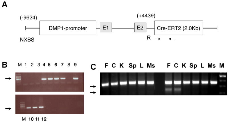
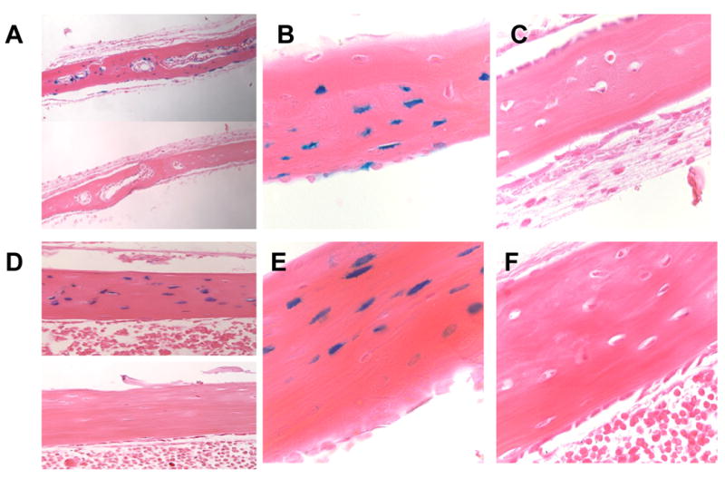
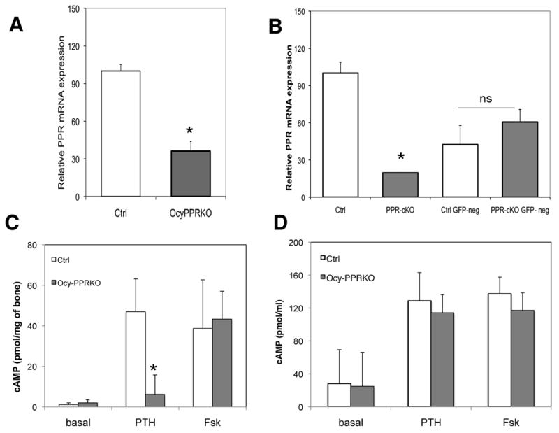
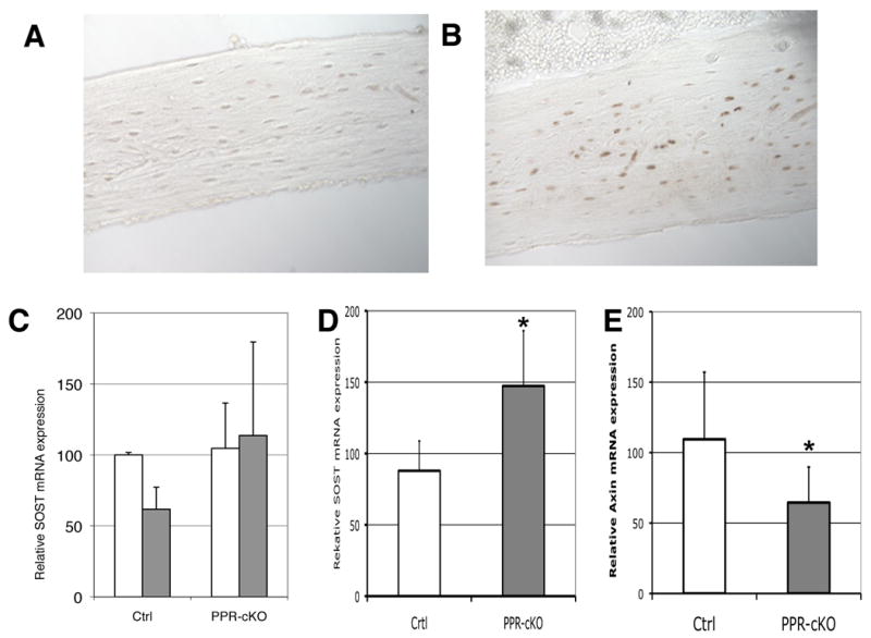
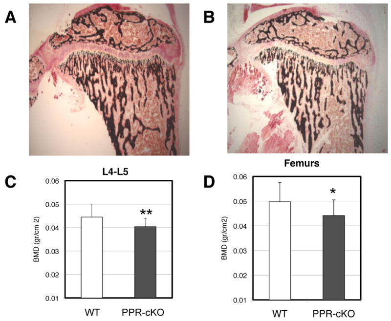
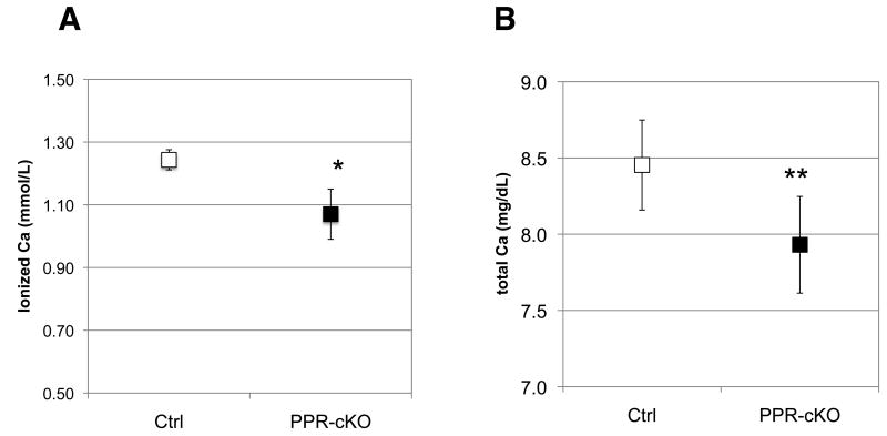
Parathyroid hormone (PTH) is a major physiologic regulator of calcium, phosphorous, and skeletal homeostasis. Cells of the osteoblastic lineage are key targets of PTH action in bone, and recent evidence suggests that osteocytes might be important in the anabolic effects of PTH. To understand the role of PTH signaling through the PTH/PTHrP receptors (PPR) in osteocytes and to determine the role(s) of these cells in mediating the effects of the hormone, we have generated mice in which PPR expression is specifically ablated in osteocytes. Transgenic mice in which the 10 kb-Dmp1 promoter drives a tamoxifen-inducible Cre-recombinase were mated with animals in which exon 1 of PPR is flanked by lox-P sites. In these animals, osteocyte-selective PPR knockout (Ocy-PPR(cKO) mice) could be induced by administration of tamoxifen. Histological analysis revealed a reduction in trabecular bone and mild osteopenia in Ocy-PPR(cKO) mice. Reduction of trabeculae number and thickness was also detected by micro-computed tomography analysis whereas bone volume fraction (BV/TV%) was unchanged. These findings were associated with an increase in Sost and sclerostin expression. When Ocy-PPR(cKO) mice were subjected to a low-calcium diet to induce secondary hyperparathyroidism, their blood calcium levels were significantly lower than littermate controls. Moreover, PTH was unable to suppress Sost and sclerostin expression in the Ocy-PPR(cKO) animals, suggesting an important role of PTH signaling in osteocytes for proper bone remodeling and calcium homeostasis.


Conflict of interest statement

Declaration of Interest: The Authors have nothing to disclose

Figures

Figure 1
Figure 1. Generation of 10KbDMP1-Cre-ERT2 mice
A) The 9.6Kb of DMP1 was cloned into the SK plasmid. NXBS and R are unique restriction sites N=NotI, X=Xbe. B=BmH, S=SmaI. R= EcoRI. The vector was released by NotI-SalI digestion. E1 and E2 are Exon 1 and the portion of Exon2 before the ATG initiation site, respectively. Arrows indicate approximate sites of PCR primers for Cre-recombinase. B) Genomic DNA from founders was assessed for integration of Cre recombinase by PCR using primers specific for the Cre-ERT2 region (arrows). Genomic DNA from the second round of pronuclear injections is shown. Eight mice were positive for Cre-recombinase (lanes 4-7, 9-12). M=100bp ladder. H = no DNA control (water). Arrows indicate the expected Cre product (size 300bp). C) Allele specific DNA recombination PCR in control (left lines) and Ocy-PPRcKO (right lines) mice. A specific band (∼ 600) is present only in femur (F) and calvaria (C) of Ocy-PPRcKO. The presence of the larger band (∼1.5 Kb) is due to contamination with other bone cells (i.e. bone marrow cells, osteoblasts and osteoclasts) that were not removed during DNA preparation. As shown here, there was no recombination in kidney (K), spleen (Sp), liver (L) or muscle (Ms). M = 100bp marker.
Figure 2
Figure 2. Characterization of DMP1-Cre-ET2 mice
X-gal staining of 5 weeks old calvarial (A-C) and long bones (femur)(D-F) from Rosa26:10KbDMP1-Cre-positive (A top panel and B: calvaria ; D top panel and E: femur) and Cre-negative (A bottom panel and C: calvaria; D bottom panel and F; femur) mice. Animals were injected with tamoxifen at days 3,5, 7, 14 and 21 days postnatal and sacrificed at 23 days (2 days after last tamoxifen injection). Panel A and D: 20X magnification; panel B,C,E and F : 40X magnification.
Figure 3
Figure 3. PPR ablation in osteocytes
Realtime qPCR from A) RNA extracted from femurs of 5 week old littermate controls (Ctrl, n=3) and Ocy-PPRcKO (n=3) mice. Data are expressed as relative mRNA, using the –ΔΔCT formula, normalized by GAPDH and expressed as percentage of controls. PPR expression in the Ocy-PPRcKO is decreased to 36.1 ± 7.7 % of controls. Data are expressed as mean ± SD of triplicates. * p<0.05. Representative experiment. B) Realtime qPCR of RNA extracted from sorted GFP-positive and GFP negative calvarial cells derived from Ocy-PPRcKO (PPR-cKO) and control littermates (Ctrl). Data are expressed as percentage of controls and are corrected for GAPDH. PPR expression in GFP-positive Ocy-PPRcKO osteocytes (PPR-KO) is decreased by 80% compared to littermate control GFP-positive osteocytes (Ctrl) (two left-most bars). PPR expression in GFP-negative cells (osteoblasts) from controls and Ocy-PPRcKO animals is not significantly different (two right-most bars). Data are expressed as mean ± SD of triplicates. * p<0.05. Representative experiment. C) Cyclic AMP accumulation in bone-marrow deprived, collaged-digested tibial explants. Right tibias of 5 week old Ocy-PPRcKO (n= 3, black bars) and control littermates (n=4, grey bars) were assayed for their in vitro responsiveness to 100nM hPTH(1-34) or 10μM forskolin (Fsk). Data are expressed as pmol of cAMP/mg of bone. Data are expressed as mean ± SD of triplicates * p<0.002 Ocy-PPRcKO vs WT. D) Cyclic AMP accumulation in primary calvaria cells. Primary calvarial cells from 4-5 days old pups were were assayed for their in vitro responsiveness to 100nM hPTH(1-34) or 10μM forskolin (Fsk). Data are expressed as pmol cAMP/well. Data are expressed as mean ± SD of triplicates.
Figure 4
Figure 4. Sost and Sclerostin expression in Ocy-PPRcKO
Immunohistochemisty for sclerostin was performed on sections of bone isolated from Ocy-PPRcKO (A) and littermate controls (B) pretreated with 0.5 mg tamoxifen 3 × week for 4 weeks prior to acute sc injection with 300μg/kg of PTH(1-34). Mice were sacrificed 1 hr after the hormone injection. C) Real-time qPCR of SOST mRNA from femurs of control littermates (Ctrl) and Ocy-PPRcKO (PPR-cKO) acutely injected with 300μg/kg of PTH(1-34)(grey bars) or vehicle alone (open bars). Data are expressed as percentage of vehicle-treated mice and normalized for GAPDH. D) Real-time qPCR of SOST mRNA from femurs of 4-6 weeks Ocy-PPRcKO (PPR-cKO n=9) and control littermates (Ctrl n=8). * p<0.005. E) Real-time qPCR of Axin-2 mRNA from femurs of 4-6 weeks Ocy-PPRcKO (PPR-cKO n=7) and control littermates (WT n=7). * p<0.05.
Figure 5
Figure 5. Osteopenia in Ocy-PPRcKO
Von Kossa (A and B) staining of 3-4 weeks old tibia of controls (A) and Ocy-PPRcKO (B) mice. Mice were injected with tamoxifen as described in Figure 2. Ocy-PPRcKO animals demonstrated a decrease in trabecular bone and a delay in secondary ossification center. C and D) Low Bone Mineral density in Ocy-PPRcKO. BMD of vertebral bodies (L4-L5)(E) and femurs (F) of Ocy-PPRcKO (n=11) and controls (n=13) females. BMD was measured by DXA on isolated bones. Data are expressed as mean ± SD. * p<0.1 ** p< 0.05.
Figure 6
Figure 6. Plasma ionized and total calcium levels in Ocy-PPRcKO(PPR-KO) and littermate controls (Ctrl) mice
Six weeks old mice were treated with 0.5 mg tamoxifen 3 times/week for 3 weeks and challenged with a low calcium diet for 2 weeks. Data are expressed as mean ± SD. Blood was collected at the end of the 3 weeks and analyzed for calcium content. Data are expressed as mean ± SD Ocy-PPRcKO n=5, controls n=3 * p<0.05. A) ionized calcium; B) total calcium.

References

    1. Aguirre JI, Plotkin LI, Stewart SA, Weinstein RS, Parfitt AM, Manolagas SC, Bellido T. Osteocyte apoptosis is induced by weightlessness in mice and precedes osteoclast recruitment and bone loss. J Bone Miner Res. 2006;21:605–615. - PubMed
    1. Ai M, Holmen SL, Van Hul W, Williams BO, Warman ML. Reduced affinity to and inhibition by DKK1 form a common mechanism by which high bone mass-associated missense mutations in LRP5 affect canonical Wnt signaling. Mol Cell Biol. 2005;25:4946–4955. - PMC - PubMed
    1. Bouxsein ML, Boyd SK, Christiansen BA, Guldberg RE, Jepsen KJ, Muller R. Guidelines for assessment of bone microstructure in rodents using micro-computed tomography. J Bone Miner Res. 25:1468–1486. - PubMed
    1. Calvi LM, Sims NA, Hunzelman JL, Knight MC, Giovannetti A, Saxton JM, Kronenberg HM, Baron R, Schipani E. Activated parathyroid hormone/parathyroid hormone-related protein receptor in osteoblastic cells differentially affects cortical and trabecular bone. J Clin Invest. 2001;107:277–286. - PMC - PubMed
    1. Cullinane DM. The role of osteocytes in bone regulation: mineral homeostasis versus mechanoreception. J Musculoskelet Neuronal Interact. 2002;2:242–244. - PubMed

Publication types

MeSH terms

Substances