. 2011 Mar 18;286(11):9677-87.
doi: 10.1074/jbc.M110.187864. Epub 2011 Jan 10.

The Ca2+ channel beta4c subunit interacts with heterochromatin protein 1 via a PXVXL binding motif

Affiliations

The Ca2+ channel beta4c subunit interacts with heterochromatin protein 1 via a PXVXL binding motif

Xingfu Xu et al. J Biol Chem. .

Abstract

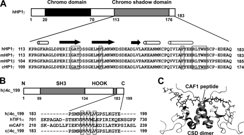
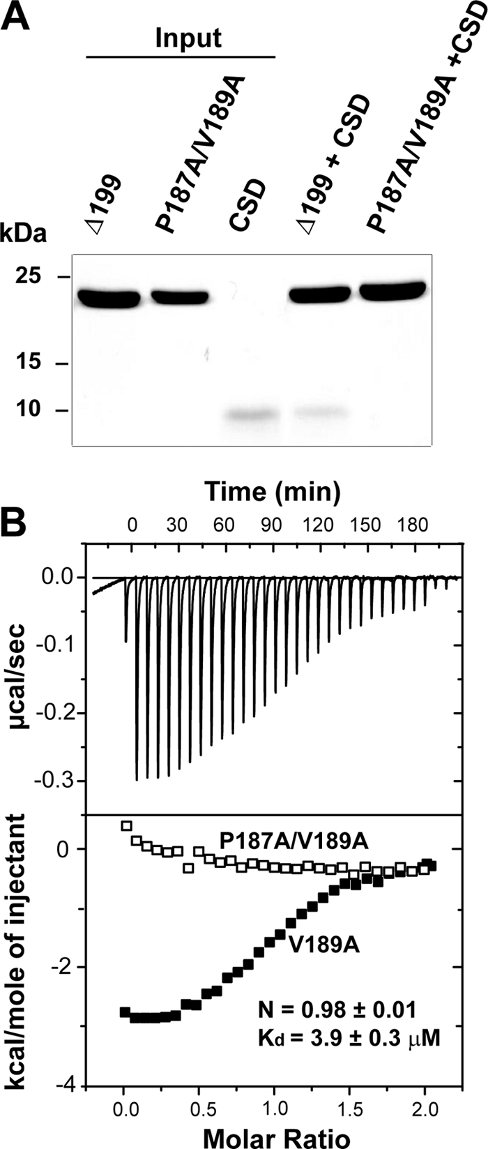
The β subunits of voltage-gated Ca(2+) channels are best known for their roles in regulating surface expression and gating of voltage-gated Ca(2+) channel α(1) subunits. Recent evidence, however, indicates that these proteins have a variety of Ca(2+) channel-independent functions. For example, on the molecular level, they regulate gene expression, and on the whole animal level, they regulate early cell movements in zebrafish development. In the present study, an alternatively spliced, truncated β4 subunit (β4c) is identified in the human brain and shown to be highly expressed in nuclei of vestibular neurons. Pull-down assays, nuclear magnetic resonance, and isothermal titration calorimetry demonstrate that the protein interacts with the chromo shadow domain (CSD) of heterochromatin protein 1γ. Site-directed mutagenesis reveals that the primary CSD interaction occurs through a β4c C-terminal PXVXL consensus motif, adding the β4c subunit to a growing PXVXL protein family with epigenetic responsibilities. These proteins have multiple nuclear functions, including transcription regulation (TIF1α) and nucleosome assembly (CAF1). An NMR-based two-site docking model of β4c in complex with dimerized CSD is presented. Possible roles for the interaction are discussed.


Figures

FIGURE 1.
FIGURE 1.
Identification of a truncated splice variant of the human Ca2+ channel β4 subunit. A, diagram of exon structure and corresponding protein domains of the full-length human β4 subunit (486 amino acids). The arrows indicate two primer sites used to detect the alternative splicing (skipping) of exon 9 (left). SH3, Src homology 3 domain. Right, results of PCR amplification using brain and cerebellum cDNA as template. The larger amplified fragment (433 bp) corresponds to a β4 product containing exon 9, whereas the smaller fragment (374 bp) corresponds to a product without exon 9. B, diagram of splice variant mRNA and resulting truncated β4c protein (left). The arrows indicate primer sites used for amplification and cloning of the coding region of the truncated splicing variant, β4c (left). Note that the downstream primer spans exons 8 and 10. The deduced domains of the human β4c protein are also shown (207 amino acids). Right, a single 639 bp band was PCR-amplified using pons cDNA as template. The cDNA codes for full-length β4c. C, sequence alignment of human and chicken β4c proteins. Tubes above the sequence indicate α-helical secondary structure. Arrows, β-strands. *, divergent amino acids. Underlined sequence delineates the β4cΔ199 construct. ∨, β4cΔ199 truncation site used to create β4cΔ184.
FIGURE 2.
FIGURE 2.
Two isoforms of the β4a subunit in mouse cerebellum and brainstem. A, Western blot showing that an affinity-purified β4a-specific polyclonal antibody labels a 52–55-kDa protein in a crude mouse cerebellar/brainstem homogenate (CB) as well as in sucrose step gradient-purified synaptosomes (S). The same antibody labels an 18–20-kDa protein in a slow speed pellet containing nuclei (N). Numbers to the left indicate molecular mass in kDa. B, distribution of β4a isoforms in cerebellar cortex and brainstem. Antibody labeling of 50-μm-thick sections was assessed by light microscopy (a, c, and e). Sections were counterstained with basic fuschin to label nuclei throughout the section (b, d, and f). Nuclear staining varies from none (a) to sparse (c) to significant (e), depending on the region of the cerebellum or brainstem. Punctate labeling of the β4a subunit can be seen in the molecular layer (ML) of the cerebellar cortex, whereas there is no specific labeling in the Purkinje (P) or granular cell (GC) layers (a). Fuschin staining confirms that the granular cell layer is rich in cell nuclei (b). c and d, sparse labeling of nuclei in the dorsal cochlear nucleus (DCN) relative to the number of fuschin-stained nuclei present. e and f, a significant number of nuclei labeled in the medial vestibular nucleus (MVN) relative to the number of fuschin-stained nuclei present. Staining of nuclei in the medial vestibular nucleus is quite variable. The predominant pattern within nuclei (inset) is suggestive of staining in heterochromatin foci.
FIGURE 3.
FIGURE 3.
The β4c protein contains a PXVXL consensus HP1 binding motif. A, chromo shadow domains from different HP1 proteins. The proteins are as follows (from top to bottom): human HP1γ, mouse HP1γ, mouse HP1β, and chicken HP1γ. CSD residues shown previously to be involved in the interaction with PXVXL-containing proteins are boxed. B, a PXVXL motif is located in the C terminus of the β4c subunit. Alignment with other PXVXL motifs from other proteins suggests its role in binding to CSD. The PXVXL-containing proteins are human Ca2+ channel β4c (hβ4c_199), human transcriptional intermediary factor 1α (hTIF1α), mouse chromatin assembly factor 1 (mCAF1), and chicken Ca2+ channel β4c (cβ4c_199). C, NMR structure of a complex (Protein Data Bank entry 1S4Z) formed by mCAF1 peptide (shown as sticks) and CSD of mouse HP1β (shown as ribbons) (20, 39). SH3, Src homology 3 domain.
FIGURE 4.
FIGURE 4.
A CSD dimer interacts specifically with β4cΔ199. A, SDS-polyacrylamide gel showing results of pull-down experiments. Lanes 1–3 contain individual proteins used in the experiments (Input): His6-β4cΔ184; His6-β4cΔ199; and untagged CSD, respectively. Lane 4 shows that when combined with β4cΔ184 beads, CSD is not present in the pellet. Lane 5 shows that when combined with β4cΔ199 beads, soluble CSD does spin down with the pellet. B, isothermal titration calorimetry. Top, raw data after base-line correction shows saturating binding of CSD when titrated into β4cΔ199 solution. Bottom, integrated data corrected for the heat of dilution of the CSD dimer. Open and filled rectangles, β4cΔ184 and β4cΔ199 data, respectively. The solid line in the bottom panel represents the best fit to a one-site binding model of the interaction of the CSD dimer with β4cΔ199.
FIGURE 5.
FIGURE 5.
NMR evidence for a specific interaction between β4cΔ199 and CSD. A, two identical overlaid HSQC spectra of 15N-labeled Δ184 in the absence (black, below) and presence (red, above) of unlabeled CSD. This is indicative of no interaction. B, two overlaid HSQC spectra of 15N-labeled β4cΔ199 in the absence (black, below) and presence (red, above) of unlabeled CSD. Note that the spectra are not identical and that many of the cross-peaks have undergone broadening or chemical shift changes that are indicative of complex formation. Residues Val189 (V189), Leu190 (L190), Gly192 (G192), and Gly197 (G197) are labeled as examples of resonances undergoing these changes.
FIGURE 6.
FIGURE 6.
Mutations in the PXVXL motif eliminate β4cΔ199-CSD dimer interaction. A, SDS-polyacrylamide gel showing results of pull-down experiments. Lanes 1–3 contain individual proteins used in the experiments: His6- β4cΔ199; His6-β4cΔ199/P187A/V189A; and untagged CSD, respectively (Input). Lane 4 shows that when combined with β4cΔ199 beads, CSD is present in the pellet. Lane 5 shows that when combined with β4cΔ199/P187A/V189A beads, soluble CSD does not spin down with the pellet. B, isothermal titration calorimetry. Top, raw data after base-line correction shows saturating binding of CSD when titrated into β4cΔ199/V189A solution. Bottom, integrated data corrected for the heat of dilution of the CSD dimer. Open and filled rectangles, β4cΔ199/P187A/V189A and β4cΔ199/V189A data, respectively. Binding data were derived from the best fit to a one-site binding model of the interaction of the CSD dimer with β4cΔ199/V189.
FIGURE 7.
FIGURE 7.
Chemical shift mapping of β4c peptide-binding region on HP1γ CSD. A, two overlaid HSQC spectra of 15N HP1γ CSD in the absence (black, above) and presence (red, below) of a 17-mer PXVXL-containing peptide derived from β4c. Residues experiencing large chemical shifts are labeled with blue lines. B, comparison of β4c binding to HP1γ CSD and CAF1 binding to HP1β CSD dimers. Residues experiencing large chemical shift changes in the complex are colored in red. Residues without assignment are shown in gray. The remainder are green. Left, mapping of the β4c peptide-binding region on the modeled structure of human HP1γ CSD dimer (see “Experimental Procedures”). The CSD dimer structure is represented in ribbons. Right, previously published mapping of mouse CAF1 peptide-binding region on the CSD dimer of mouse HP1β (39).
FIGURE 8.
FIGURE 8.
Two sites of β4cΔ199 interaction with CSD based on modeling and NMR. A, HADDOCK (high ambiguity-driven docking) (21) model of the complex of β4cΔ199 (purple ribbon) with the CSD dimer (green ribbons). The β4cΔ199 C terminus with the PXVXL binding motif is shown in orange. B, view of part of the overlaid spectra of 15N-labeled CSD in the absence (black, below) and in the presence (red, above) of β4cΔ199. The highlighted CSD residues undergoing chemical shifts (Glu179–Gln183; 179E–183Q) are from one monomer of the CSD dimer in complex with β4cΔ199.

References

    1. Dolphin A. C. (2003) J. Bioenerg. Biomembr. 35, 599–620 - PubMed
    1. Dolphin A. C. (2009) Curr. Opin. Neurobiol. 19, 237–244 - PubMed
    1. De Waard M., Pragnell M., Campbell K. P. (1994) Neuron 13, 495–503 - PubMed
    1. Ebert A. M., McAnelly C. A., Srinivasan A., Linker J. L., Horne W. A., Garrity D. M. (2008) Proc. Natl. Acad. Sci. U.S.A. 105, 198–203 - PMC - PubMed
    1. Subramanyam P., Obermair G. J., Baumgartner S., Gebhart M., Striessnig J., Kaufmann W. A., Geley S., Flucher B. E. (2009) Channels 3, 343–355 - PMC - PubMed

Publication types

MeSH terms