. 2011 Mar 11;286(10):8577-8584.
doi: 10.1074/jbc.M110.187179. Epub 2011 Jan 10.

Cooperativeness of Orai cytosolic domains tunes subtype-specific gating

Affiliations

Cooperativeness of Orai cytosolic domains tunes subtype-specific gating

Irene Frischauf et al. J Biol Chem. .

Abstract

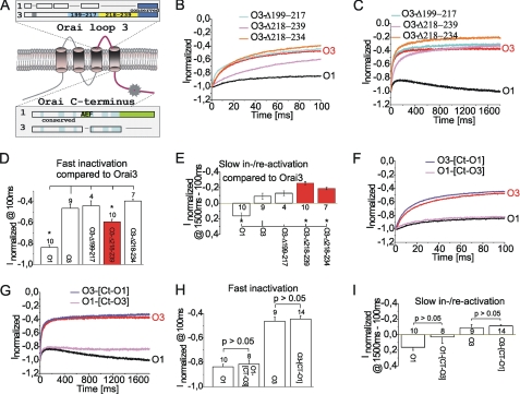
Activation of immune cells is triggered by the Ca(2+) release-activated Ca(2+) current, which is mediated via channels of the Orai protein family. A key gating process of the three Orai channel isoforms to prevent Ca(2+) overload is fast inactivation, most pronounced in Orai3. A subsequent reactivation is a unique gating characteristic of Orai1 channels, whereas Orai2 and Orai3 currents display a second, slow inactivation phase. Employing a chimeric approach by sequential swapping of respective intra- and extracellular regions between Orai1 and Orai3, we show here that Orai1 specific proline/arginine-rich domains in the N terminus mediate reactivation, whereas the second, intracellular loop modulates fast and slow gating processes. Swapping C-terminal strands lacks a significant impact. However, simultaneous transfer of Orai3 N terminus and its second loop or C terminus in an Orai1 chimera substantially increases fast inactivation centered between wild-type channels. Concomitant swap of all three cytosolic strands from Orai3 onto Orai1 fully conveys Orai3-like gating characteristics, in a strongly cooperative manner. In conclusion, Orai subtype-specific gating requires a cooperative interplay of all three cytosolic domains.


Figures

FIGURE 1.
FIGURE 1.
The proline/arginine-rich regions of the Orai1 N terminus mediate reactivation. A, a scheme depicts an Orai protein with its four transmembrane segments and highlights the unique arginine/proline/arginine-rich region within Orai1. R, Arg; P, Pro. B and C, normalized average currents from a voltage step to −82 mV are shown for a coexpression of STIM1 with Orai1 (O1, black), Orai3 (O3, red), as well as O1-[Nt-O3], O3-[Nt-O1], or O1-Δ1–47 for 100 (B) or 1800 (C) ms. D, normalized average currents were calculated for a time point at 100 ms representing fast inactivation (blue arrows) and slow inactivation/reactivation (cyan arrows) was estimated by subtracting normalized currents at 1500 ms from those at 100 ms. E and F, statistics on normalized average currents ± S.E. compare O1, O1-Δ1–38, O1-Δ1–47, O1-[NT-O3], O3-[NT-O1], O3, and O3-[ins27–51-O1] at 100 (E) and 1500–100 ms (F). G, time course of average FRET change ± S.E. between CFP-CaM or CFP-CaMMUT and YFP-O1-Nt or YFP-O3-Nt in response to addition of 10 μm ionomycin (Iono) and 2 mm Ca2+ in a bath solution administered at 1 min. At 3 min, the relative FRET values of YFP-O1-Nt or YFP-O3-Nt coexpressed with CFP-CAM is significantly different (p < 0.01) to a coexpression of either Orai-Nt with the CaMMUT. H and I, localization, overlay, and calculated FRET life cell image series of CFP-CaM and YFP-Orai1-Nt (H) or YFP-Orai3-Nt (I) in a nominally Ca2+ extracellular solution (upper images) or with 2 mm Ca2+ and 10 μm ionomycin added (lower images).
FIGURE 2.
FIGURE 2.
The second loop of Orai1 transferred into Orai3 reduces fast inactivation. A, the scheme depicts an Orai protein and highlights nonconserved residues within the second loop of Orai1 or Orai3. B and C, normalized average currents from a voltage step to −82 mV are shown for a coexpression of STIM1 with O1 (black), O3 (red), as well as O1-[L2-O3] and O3-[L2-O1] for 100 (B) or 1800 (C) ms. D and E, statistics on normalized average currents ± S.E. compare O1, O1-[L2-O3], O3, and O3-[L2-O1] at 100 (D) and 1500–100 (E) ms.
FIGURE 3.
FIGURE 3.
Effect of the third loop and the C terminus on Orai gating. A, the scheme depicts an Orai protein and highlights unique insertions within the third loop of Orai3 as well as C terminus of Orai1. B and C, normalized average currents from a voltage step to −82 mV are shown for a coexpression of STIM1 with O1 (black), O3 (red), as well as O3-Δ199–217, O3-Δ218–239, and O3-Δ218–234 for 100 (B) or 1800 (C) ms. Statistics on normalized average currents ± S.E. compare O1, O3, O3-Δ199–217, O3-Δ218–239, and O3-Δ218–234 at 100 (D) and 1500–100 (E) ms. F–I, analogous voltage steps over 100 (F) or 1800 (G) ms and statistics on average currents ± S.E. at 100 (H) or 1800 (I) ms are presented for coexpression of STIM1 with O1, O3, as well as O1-[Ct-O3] and O3-[Ct-O1].
FIGURE 4.
FIGURE 4.
Orai1 chimera with two Orai3 domains swaps exhibit increased fast inactivation. A and B, normalized average currents from a voltage step to −82 mV are shown for a coexpression of STIM1 with O1 (black), O3 (red), as well as O1-[Nt-O3], O1-[Ct-O3], and O1-[L2-O3] for 100 (A) or 1800 (B) ms. Analogous voltage steps are presented for either O1-[Nt-Ct-O3], O1-[Nt-L2-O3], and O1-[Nt-L2-Ct-O3] for 100 (C) or 1800 (D) ms. Statistics on normalized average currents ± S.E. are compared for significant difference for O1 to O1-[Nt-L2-O3], O1-[Nt-Ct-O3], O1-[Nt-L2-Ct-O3], and O3 at 100 (E) and 1500–100 (F) ms. An addition of normalized average currents of O1-[Nt-O3], O1-[L2-O3], and O1-[Ct-O3] are shown at 100 ms included in E.
FIGURE 5.
FIGURE 5.
Orai3 chimera with two Orai1 domains swaps show reduced fast inactivation. A and B, normalized average currents from a voltage step to −82 mV are shown for a coexpression of STIM1 with O1 (black), O3 (red), as well as O3-[Nt-O1], O3-[L2-O1], and O3-[Ct-O1] for 100 (A) or 1800 (B) ms. Analogous voltage steps are presented for either O3-[Nt-L2-O1], O3-[Nt-Ct-O1], and O3-[L2-Ct-O1] for 100 (C) or 1800 (D) ms. Statistics on normalized average currents ± S.E. of O1, O3-[Nt-Ct-O1], O3-[Nt-L2-O1], and O3-[Ct-L2-O1] are compared for significant difference with O3 at 100 (E) and 1500–100 (F) ms.
FIGURE 6.
FIGURE 6.
Gating of Orai1 and Orai3 chimera with identical combinations of cytosolic strands. A, normalized average currents from a voltage step to −82 mV are shown for a coexpression of STIM1 with O1 (black), O3 (red), as well as O1-[Nt-O1] and O3-[L2-Ct-O1] for 1800 ms. Analogous voltage steps compare O1-[L2-Ct-O3] and O3-[Nt-O1] (B), O1-[L2-O3] and O3-[Nt-Ct-O1] (C), O1-[Nt-Ct-O3] and O3-[L2-O1] (D), O1-[Ct-O3] and O3-[Nt-L2-O1] (E) as well as O1-[Nt-L2-O3] and O3-[Ct-O1] (F).

References

    1. Parekh A. B., Putney J. W., Jr. (2005) Physiol. Rev. 85, 757–810 - PubMed
    1. Roos J., DiGregorio P. J., Yeromin A. V., Ohlsen K., Lioudyno M., Zhang S., Safrina O., Kozak J. A., Wagner S. L., Cahalan M. D., Veliçelebi G., Stauderman K. A. (2005) J. Cell Biol. 169, 435–445 - PMC - PubMed
    1. Liou J., Kim M. L., Heo W. D., Jones J. T., Myers J. W., Ferrell J. E., Jr., Meyer T. (2005) Curr. Biol. 15, 1235–1241 - PMC - PubMed
    1. Luik R. M., Wang B., Prakriya M., Wu M. M., Lewis R. S. (2008) Nature 454, 538–542 - PMC - PubMed
    1. Muik M., Frischauf I., Derler I., Fahrner M., Bergsmann J., Eder P., Schindl R., Hesch C., Polzinger B., Fritsch R., Kahr H., Madl J., Gruber H., Groschner K., Romanin C. (2008) J. Biol. Chem. 283, 8014–8022 - PubMed

Publication types

LinkOut - more resources