. 2011 Mar;17(3):535-43.
doi: 10.1261/rna.2436411. Epub 2011 Jan 10.

A quantitative assay for measuring mRNA decapping by splinted ligation reverse transcription polymerase chain reaction: qSL-RT-PCR

Affiliations

A quantitative assay for measuring mRNA decapping by splinted ligation reverse transcription polymerase chain reaction: qSL-RT-PCR

Nathan Blewett et al. RNA. 2011 Mar.

Abstract

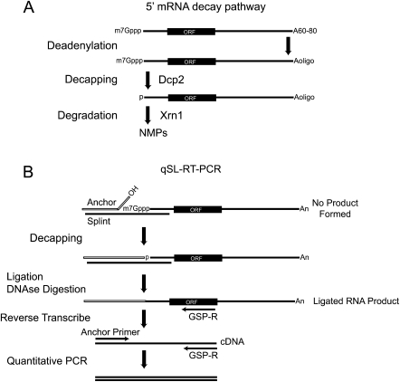
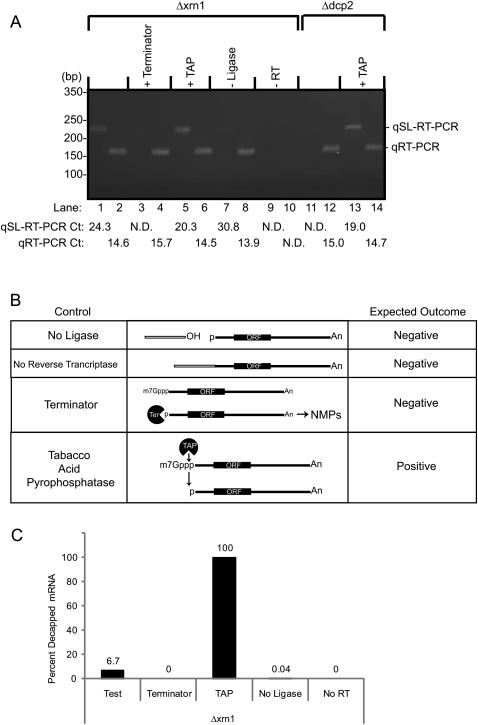
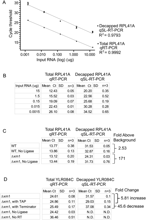
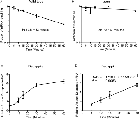
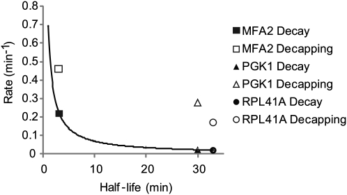
The degradation of messenger RNA is a critical node of gene regulation. A major pathway of mRNA decay is initiated by shortening of the poly(A) tail, followed by removal of the 5' cap structure (decapping) and subsequent degradation. Decapping is an important determinate in the destruction of many transcripts. Detailed kinetic analysis of in vivo decapping rates is necessary to understand how this step is regulated. Importantly, the product of decapping is recalcitrant for investigation, in part due to its transient nature. As such, little in vivo kinetic information is available. Here we report the development of an assay that measures decapping of mRNAs by combining splinted ligation and quantitative RT-PCR (qSL-RT-PCR). We apply this method to determine the decapping rate constant for a natural mRNA in vivo for the first time. The qSL-RT-PCR assay may be adapted for use on any mRNA, providing a new tool to study regulation of mRNA decay.


Figures

FIGURE 1.
FIGURE 1.
The 5′ mRNA decay pathway. (A) mRNAs possess a 5′ 7-methyl guanosine cap (7mGppp), open reading frame (ORF) and a 3′ poly-adenosine tail, averaging 60–80 nt in yeast. mRNA decay via the 5′ pathway initiates with deadenylation of the poly-Adenosine tail to a short oligo-adenylated form (Aoligo). Dcp2 decapping enzyme subsequently removes the cap. The decapped mRNA, with a 5′ monophosphate, is destroyed by Xrn1, producing monophosphorylated nucleotides (NMP). (B) Schematic of the quantitative splinted-ligation reverse transcriptase polymerase chain reaction assay (qSL-RT-PCR). First, Anchor RNA and complementary DNA Splint oligonucleotides are annealed to the 5′ end of the target mRNA. mRNAs containing a 5′ cap cannot be ligated, as the cap prevents ligation. Decapped RNAs have a 5′ monophosphate that is ligated to the Anchor RNA 3′ hydroxyl by T4 DNA ligase. After ligation, the splint is destroyed by DNase I. The ligated RNA is converted to cDNA by reverse transcription with a reverse gene-specific primer (GSP-R). The resulting cDNA is then detected by quantitative PCR using GSP-R and a forward primer that anneals to the anchor (Anchor Primer). An internal control qPCR is performed on the same cDNA samples using gene-specific primers that amplify within the coding sequence of the mRNA.
FIGURE 2.
FIGURE 2.
The qSL-RT-PCR assay specifically detects decapped mRNA. (A) qSL-RT-PCR was used to detect endogenous decapped RPL41A mRNA present in 10 μg of total RNA isolated from yeast strains lacking either the XRN1 (Δxrn1) or the DCP2 (Δdcp2) genes (odd-numbered lanes). Standard qRT-PCR was used to detect total RPL41A RNA (even-numbered lanes). In control reactions, total RNA was treated as indicated at the top of the gel, including Terminator (lanes 3,4) or Tobacco Acid Pyrophosphatase (+TAP) treatment (lanes 5,6,13,14). In lanes 7 and 8, DNA ligase was omitted (−Ligase). Reverse transcriptase was omitted from reactions in lanes 9 and 10 (–RT). Cycle thresholds measured for each sample are indicated at the bottom of the figure. Reactions that did not yield a detectable cycle threshold are labeled “N.D.” (Not Detected). (B) Critical controls for the qSL-RT-PCR assay demonstrate specificity for decapped mRNA. Control reactions are indicated on the left. In the middle, a diagram of each control is depicted. On the right, the expected outcome of qSL-RT-PCR is indicated for each control reaction. (Terminator) The Terminator enzyme specifically destroys uncapped mRNA with a 5′ monophosphate but does not degrade capped mRNA, thereby demonstrating specificity of the assay for decapped RNA. Tobacco Acid Phosphatase removes 5′ cap, leaving a 5′ monophosphate that is detected by the qSL-RT-PCR assay, thereby serving as a positive control. (No ligase) Omission of DNA ligase prevents ligation of anchor to mRNA, thereby preventing amplification of product. (No Reverse Transcriptase) In the absence of reverse transcriptase, no product should be generated, thereby demonstrating dependence on cDNA conversion of mRNA. (C) Relative amounts of decapped RPL41A mRNA in A were determined for the indicated reactions from the strain lacking Xrn1. Calculations are described in the Materials and Methods.
FIGURE 3.
FIGURE 3.
The qSL-RT-PCR assay has a broad, linear dynamic range for sensitive detection of decapped RPL41A RNA. (A) Five 10-fold serial dilutions of total RNA from Δxrn1 cells were analyzed using qSL-RT-PCR and qRT-PCR assays to detect decapped and total RPL41A mRNA, respectively. Triplicate samples were analyzed and the mean cycle threshold (Ct) values are plotted against input RNA amount (15, 1.5, 0.15, 0.015, and 0.0015 μg) on a logarithmic scale. Standard deviation is indicated above and below each data point. (B) Mean Ct values and standard deviations (SD) from A are shown in the table. Nonlinear regression analysis was used to determine correlation coefficients (R2) for each curve. (C) qRT-PCR and qSL-RT-PCR assays were performed on 10 μg of total RNA to measure RPL41A mRNA in wild-type BY4742 cells (WT) and Δxrn1. Mean Ct values and standard deviations are indicated as determined from triplicate samples. Fold increase above background was calculated relative to control reactions lacking T4 DNA ligase for each strain, determined from ΔCt of qSL-RT-PCR. (D) qRT-PCR and qSL-RT-PCR assays were performed on 7.5 μg of total RNA to measure YLR084C mRNA in Δxrn1 cells. Control samples were treated with TAP or Terminator. T4 DNA ligase or Reverse Transcriptase were omitted from control reactions as indicated. Mean Ct values and standard deviations are indicated and were determined from triplicate samples. Fold change in decapped mRNA level was calculated from the ΔCt of qSL-RT-PCR values. Reactions that did not yield a detectable cycle threshold are labeled “N.D.” (Not Detected).
FIGURE 4.
FIGURE 4.
The in vivo decapping rate of RPL41A mRNA was determined using qSL-RT-PCR assay. The half-life of RPL41A was measured in wild-type yeast (A) and in a strain wherein the XRN1 gene is deleted (Δxrn1) (B). (C) Decapped RPL41A mRNA was measured using the qSL-RT-PCR assay in the Δxrn1 strain following transcription shutoff. Relative amount of decapped RNA was calculated after normalization to total RPL41A at each time-point, relative to time = 0. (D) The rate of decapping was determined by linear regression analysis of the data in the graph in C with linear reaction kinetics between 5 and 30 min. The slope of the line, the reaction rate, is shown along with error and correlation coefficient. In all graphs, the mean value of the replicates is plotted and standard error is indicated above and below the data points.
FIGURE 5.
FIGURE 5.
Comparison of mRNA half-life, decay, and decapping reaction rates. The reaction rates of decay (in black) and decapping (in white) are compared with mRNA half-lives of three mRNAs, MFA2, PGK1, and RPL41A. Decay rates were derived from each mRNA's half-life. The trend-line curve plots the decay rate for mRNAs with half-lives ranging from 1 to 33 min. Decapping rates and mRNA half-lives for PGK1 and MFA2 were measured previously by Northern blot analysis of the poly(G) tract containing RNA (Muhlrad and Parker 1992; Muhlrad et al. 1995; Cao and Parker 2001). RPL41A decapping rate and half-life were measured by qSL-RT-PCR and qRT-PCR, respectively, as shown in Figure 4.

References

    1. Anderson JS, Parker RP 1998. The 3′ to 5′ degradation of yeast mRNAs is a general mechanism for mRNA turnover that requires the SKI2 DEVH box protein and 3′ to 5′ exonucleases of the exosome complex. EMBO J 17: 1497–1506 - PMC - PubMed
    1. Araki Y, Takahashi S, Kobayashi T, Kajiho H, Hoshino S, Katada T 2001. Ski7p G protein interacts with the exosome and the Ski complex for 3′-to-5′ mRNA decay in yeast. EMBO J 20: 4684–4693 - PMC - PubMed
    1. Beelman CA, Stevens A, Caponigro G, LaGrandeur TE, Hatfield L, Fortner DM, Parker R 1996. An essential component of the decapping enzyme required for normal rates of mRNA turnover. Nature 382: 642–646 - PubMed
    1. Behm-Ansmant I, Rehwinkel J, Doerks T, Stark A, Bork P, Izaurralde E 2006. mRNA degradation by miRNAs and GW182 requires both CCR4:NOT deadenylase and DCP1:DCP2 decapping complexes. Genes Dev 20: 1885–1898 - PMC - PubMed
    1. Brown JT, Bai X, Johnson AW 2000. The yeast antiviral proteins Ski2p, Ski3p, and Ski8p exist as a complex in vivo. RNA 6: 449–457 - PMC - PubMed

Publication types

MeSH terms