Comparative Study
. 2011 Jan;68(1):74-84.
doi: 10.1001/archneurol.2010.342.

Deficits in functional connectivity of hippocampal and frontal lobe circuits after traumatic axonal injury

Affiliations
Comparative Study

Deficits in functional connectivity of hippocampal and frontal lobe circuits after traumatic axonal injury

Carlos D Marquez de la Plata et al. Arch Neurol. 2011 Jan.

Abstract

Objective: To examine the functional connectivity of hippocampal and selected frontal lobe circuits in patients with traumatic axonal injury (TAI).

Design: Observational study.

Setting: An inpatient traumatic brain injury unit. Imaging and neurocognitive assessments were conducted in an outpatient research facility.

Participants: Twenty-five consecutive patients with brain injuries consistent with TAI and acute subcortical white matter abnormalities were studied as well as 16 healthy volunteers of similar age and sex.

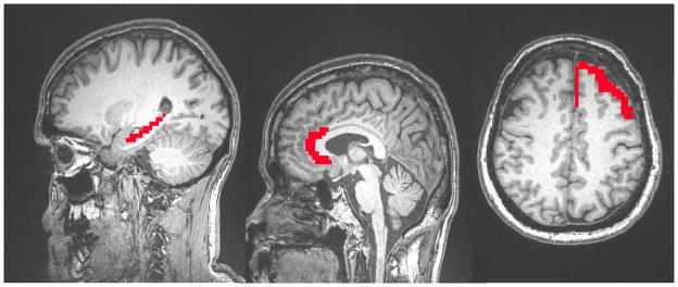
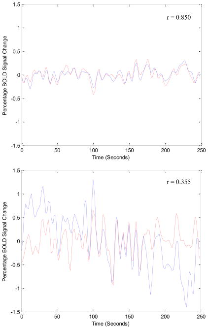
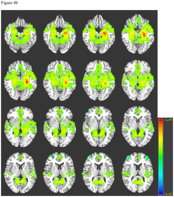
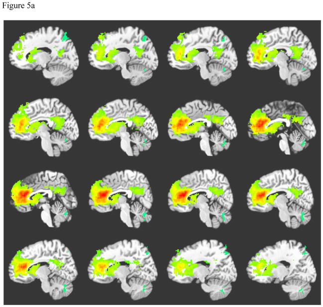
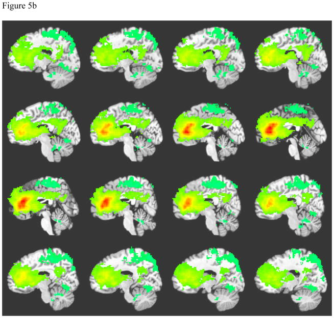
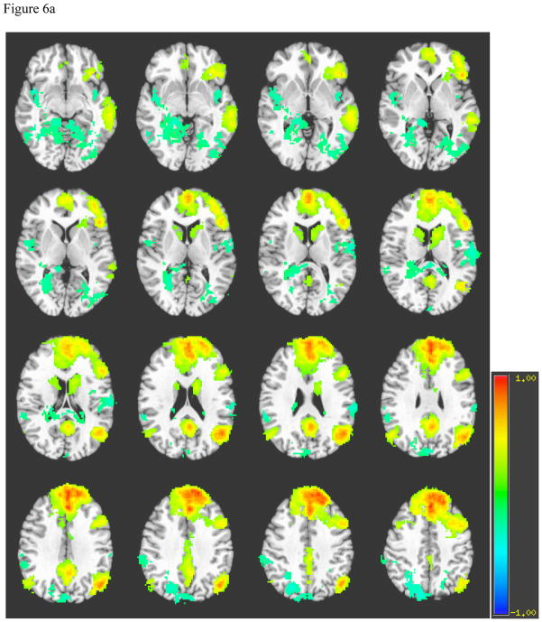
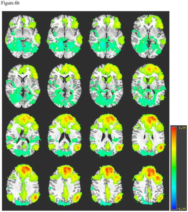
Interventions: Echo-planar and high-resolution T1-weighted images were acquired using 3-T scanners. Regions of interest (ROI) were drawn bilaterally for the hippocampus, anterior cingulate cortex (ACC), and dorsolateral prefrontal cortex and were used to extract time series data. Blood oxygenation level-dependent data from each ROI were used as reference functions for correlating with all other brain voxels. Interhemispheric functional connectivity was assessed for each participant by correlating homologous regions using a Pearson correlation coefficient. Patient functional and neurocognitive outcomes were assessed approximately 6 months after injury.

Main outcome measures: Interhemispheric functional connectivity, spatial patterns of functional connectivity, and associations of connectivity measures with functional and neurocognitive outcomes.

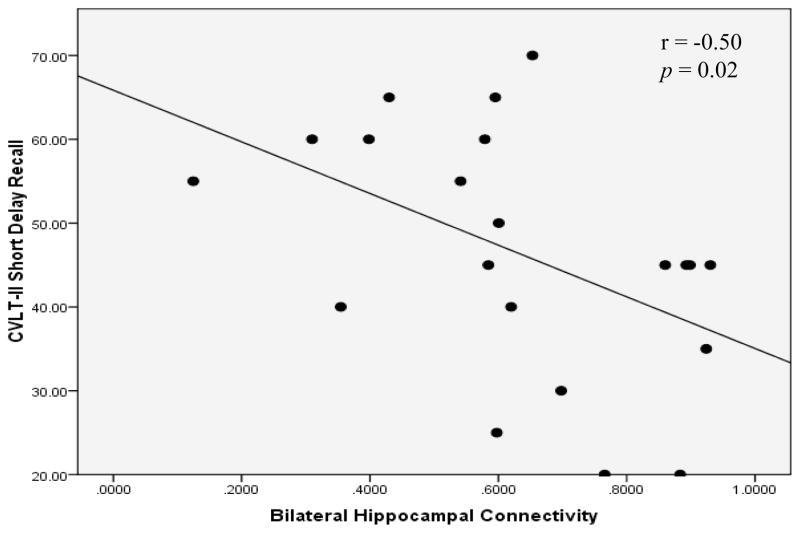
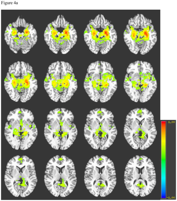
Results: Patients showed significantly lower interhemispheric functional connectivity for the hippocampus and ACC. Controls demonstrated stronger and more focused functional connectivity for the hippocampi and ACC, and a more focused recruitment of the default mode network for the dorsolateral prefrontal cortex ROI. The interhemispheric functional connectivity for the hippocampus was correlated with delayed recall of verbal information.

Conclusions: Traumatic axonal injury may affect interhemispheric neural activity, as patients with TAI show disrupted interhemispheric functional connectivity. More careful investigation of interhemispheric connectivity is warranted, as it demonstrated a modest association with outcome in chronic TBI.


Figures

Figure 1
Figure 1
Location of Reference Seed Regions of Interest. Note. Location of manually drawn regions of interest. A. Hippocampus. B. Anterior cingulate cortex. C. Left Dorsolateral Prefrontal cortex (radiologic convention).
Figure 2
Figure 2
Bilateral Hippocampal BOLD Fluctuations Over Time. Note. Interhemispheric functional connectivity for left and right hippocampi for a healthy control (above), and for a patient with TAI (below). BOLD signal change is plotted over time. Notice the BOLD signal fluctuates significantly more synchronously in a healthy brain as opposed to an injured brain.
Figure 3
Figure 3
Association between Interhemispheric Hippocampal Connectivity and Verbal Memory Outcome. Note. A Spearman correlation coefficient shows a negative association between interhemispheric hippocampal connectivity and delayed verbal memory. CVLT-II = California Verbal Learning Test-Second Edition.
Figure 4
Figure 4
Figure 4a. Average Left Hippocampus Connectivity among Normal Controls Note for Figures 4a and 4b. A= Hippocampi, B= Basal forebrain, C= Frontal Lobe, D= Hypothalamus, E= Temporal lobe, F= Septal nuclei, G= Subthalamic nuclei, H= parahippocampal gyrus, I= Posterior cingulate. Figure 4b. Average Left Hippocampus Connectivity among Patients with TAI Note: A= Hippocampi, B= Basal forebrain, C= Frontal Lobe, D= Hypothalamus, E= Temporal lobe, F= Septal nuclei, G= Subthalamic nuclei, H= parahippocampal gyrus, I= Posterior cingulate cortex.
Figure 4
Figure 4
Figure 4a. Average Left Hippocampus Connectivity among Normal Controls Note for Figures 4a and 4b. A= Hippocampi, B= Basal forebrain, C= Frontal Lobe, D= Hypothalamus, E= Temporal lobe, F= Septal nuclei, G= Subthalamic nuclei, H= parahippocampal gyrus, I= Posterior cingulate. Figure 4b. Average Left Hippocampus Connectivity among Patients with TAI Note: A= Hippocampi, B= Basal forebrain, C= Frontal Lobe, D= Hypothalamus, E= Temporal lobe, F= Septal nuclei, G= Subthalamic nuclei, H= parahippocampal gyrus, I= Posterior cingulate cortex.
Figure 5
Figure 5
Figure 5a. Average Left Anterior Cingulate Connectivity among Normal Controls Note: Crosshairs were placed on slice closest to midline. A = Right caudate, B= Anterior cingulate cortex, C= Cingulate cortex, D= Posterior cingulate cortex, E = Thalamus, F= Left caudate. Figure 5b: Average Left Anterior Cingulate Connectivity among Patients with TAI Note: Crosshairs were placed on slice closest to midline. A = Right caudate, B= Anterior cingulate cortex, C= Cingulate cortex, D = Posterior cingulate cortex, E = Thalamus, F= Left caudate.
Figure 5
Figure 5
Figure 5a. Average Left Anterior Cingulate Connectivity among Normal Controls Note: Crosshairs were placed on slice closest to midline. A = Right caudate, B= Anterior cingulate cortex, C= Cingulate cortex, D= Posterior cingulate cortex, E = Thalamus, F= Left caudate. Figure 5b: Average Left Anterior Cingulate Connectivity among Patients with TAI Note: Crosshairs were placed on slice closest to midline. A = Right caudate, B= Anterior cingulate cortex, C= Cingulate cortex, D = Posterior cingulate cortex, E = Thalamus, F= Left caudate.
Figure 6
Figure 6
Figure 6a. Average Left Dorsolateral Prefrontal Cortex Connectivity among Normal Controls Note. A = Parahippocampal gyrus, B = Occipito-temporal gyrus, C = Middle temporal gyrus, D = Inferior frontal gyrus, E = Caudate, F = Posterior cingulate cortex, G = Anterior cingulate cortex, H = Angular gyrus, I = Superior frontal gyrus, J = Precuneus. Figure 6b. Average Left Dorsolateral Prefrontal Cortex Connectivity among Patients with TAI Note. A = Parahippocampal gyrus, B = Occipito-temporal gyrus, C = Middle temporal gyrus, D = Inferior frontal gyrus, E = Caudate, F = Posterior cingulate cortex, G = Anterior cingulate cortex, H = Angular gyrus, I = Superior frontal gyrus, J = Precuneus.
Figure 6
Figure 6
Figure 6a. Average Left Dorsolateral Prefrontal Cortex Connectivity among Normal Controls Note. A = Parahippocampal gyrus, B = Occipito-temporal gyrus, C = Middle temporal gyrus, D = Inferior frontal gyrus, E = Caudate, F = Posterior cingulate cortex, G = Anterior cingulate cortex, H = Angular gyrus, I = Superior frontal gyrus, J = Precuneus. Figure 6b. Average Left Dorsolateral Prefrontal Cortex Connectivity among Patients with TAI Note. A = Parahippocampal gyrus, B = Occipito-temporal gyrus, C = Middle temporal gyrus, D = Inferior frontal gyrus, E = Caudate, F = Posterior cingulate cortex, G = Anterior cingulate cortex, H = Angular gyrus, I = Superior frontal gyrus, J = Precuneus.

References

    1. Thurman DJ, Alverson C, Dunn KA, Guerrero J, Sniezek JE. Traumatic brain injury in the United States: a public health perspective. J Head Trauma Rehabil. 1999;14(6):602–15. - PubMed
    1. Langlois M, Cramer KM. The relationship between household composition and retirement stress. Guidance & Counseling. 2004;20(1):89–98.
    1. Adams JH, Doyle D, Ford I, Gennarelli TA, Graham DI, McLellan DR. Diffuse axonal injury in head injury: Definition, diagnosis and grading. Histopathology. 1989;15(1):49–59. - PubMed
    1. Meythaler JM, Peduzzi JD, Eleftheriou E, Novack TA. Current concepts: Diffuse axonal injury-associated traumatic brain injury. Arch Phys Med Rehabil. 2001;82(10):1461–71. - PubMed
    1. Mittl RL, Grossman RI, Hiehle JF, et al. Prevalence of MR evidence of diffuse axonal injury in patients with mild head injury and normal head CT findings. AJNR, Am J Neuroradiology. 1994;15(8):1583–89. - PMC - PubMed

Publication types

MeSH terms