. 2011 Mar 17;117(11):3045-52.
doi: 10.1182/blood-2010-07-294751. Epub 2011 Jan 10.

Recombinant erythroid Kruppel-like factor fused to GATA1 up-regulates delta- and gamma-globin expression in erythroid cells

Affiliations

Recombinant erythroid Kruppel-like factor fused to GATA1 up-regulates delta- and gamma-globin expression in erythroid cells

Jianqiong Zhu et al. Blood. .

Abstract

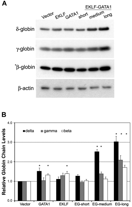
The β-hemoglobinopathies sickle cell disease and β-thalassemia are among the most common human genetic disorders worldwide. Hemoglobin A2 (HbA2, α₂δ₂) and fetal hemoglobin (HbF, α₂γ₂) both inhibit the polymerization of hemoglobin S, which results in erythrocyte sickling. Expression of erythroid Kruppel-like factor (EKLF) and GATA1 is critical for transitioning hemoglobin from HbF to hemoglobin A (HbA, α₂β₂) and HbA2. The lower levels of δ-globin expression compared with β-globin expression seen in adulthood are likely due to the absence of an EKLF-binding motif in the δ-globin proximal promoter. In an effort to up-regulate δ-globin to increase HbA2 expression, we created a series of EKLF-GATA1 fusion constructs composed of the transactivation domain of EKLF and the DNA-binding domain of GATA1, and then tested their effects on hemoglobin expression. EKLF-GATA1 fusion proteins activated δ-, γ-, and β-globin promoters in K562 cells, and significantly up-regulated δ- and γ-globin RNA transcript and protein expression in K562 and/or CD34(+) cells. The binding of EKLF-GATA1 fusion proteins at the GATA1 consensus site in the δ-globin promoter was confirmed by chromatin immunoprecipitation assay. Our studies demonstrate that EKLF-GATA1 fusion proteins can enhance δ-globin expression through interaction with the δ-globin promoter, and may represent a new genetic therapeutic approach to β-hemoglobinopathies.


Figures

Figure 1
Figure 1
Schematic diagram of the structure of human hemoglobin reporter constructs and EKLF, GATA1, and EKLF-GATA1 fusion constructs. (A) Comparison of the δ- and β-globin promoter proximal regions; GATA1- and EKLF-binding sites are indicated. (B) The β-globin locus on human chromosome 11. Schematic diagram of δ-, β-, and γ-globin promoter regions in a luciferase reporter construct with the known functional EKLF- or GATA1-binding sites in each promoter indicated.,,– (C) Schematic diagram of the structure of EKLF, GATA1, and 3 different EKLF-GATA1 fusion pLenti V5 topo expression constructs. All inserts were fused to a V5 epitope at the C-terminus with the transactivation and DNA-binding domains indicated. F1, F2, and F3 represent 3 finger domains of EKLF; CF and NF represent C- and N-fingers of GATA1; R1, R2, and R3 represent 3 regions of the transactivation domain of GATA1. (D) Long (L), medium (M), or short (S) form of EKLF, GATA1, EKLF-GATA1, or vector only (V) were transiently transfected into 293FT cells and then harvested 48 hours after transfection and subjected to Western blotting analysis using anti-V5, EKLF, or GATA1 antibodies. Mo indicates mock-transfected 293FT cells.
Figure 2
Figure 2
EKLF-GATA1 fusion proteins activated δ-, γ-, and β-globin promoter activity in K562 cells. ELKF, GATA1, fusion EKLF-GATA1 (EG) vector, or vector only was cotransfected with δ-, γ-, or β-globin promoter reporter constructs (A) or wild-type or mutant δ-globin promoter reporter constructs (B) into K562 cells. The level of promoter activity was evaluated 48 hours after transfection by measurement of firefly luciferase activity relative to the internal control renilla luciferase activity using the dual luciferase assay system essentially as described by the manufacturer. (A) Fold increase was calculated compared with expression in mock-transfected K562 cells. *P < .05 versus mock-transfected cells. (B) Fold increase was calculated relative to expression in mock-transfected cells. **P < .01 or ***P < .001 versus corresponding wild-type transfected cells. Error bars indicate SD of the mean of 3 independent experiments.
Figure 3
Figure 3
The EKLF-GATA1 fusion proteins enhanced δ-globin gene expression in K562 and CD34+ cells. (A) K562 cells were transfected with EKLF, GATA1, fusion EKLF-GATA1 (EG), or vector only and cultured in medium with 6 μg/mL of blasticidin for 2 weeks. RNA was isolated and real-time PCR analysis performed. Data represent real-time PCR analysis of δ- and γ-globin gene expression in EKLF-, GATA1-, or fusion EKLF-GATA1–transfected cells normalized to β-actin gene expression. Fold increase was calculated compared with expression in mock-transfected cells. *P < .05 versus mock-transfected cells. Error bars indicate SD of the mean of 3 independent experiments. (B) CD34+ bone marrow cells were infected by lentivirus encoding EKLF, GATA1, fusion EKLF-GATA1 (EG) constructs, or vector only at day 4 of the expansion stage, On day 6, gene-transduced CD34+ cells were reseeded and grown in differentiation medium with 3 μg/mL of blasticidin for 7 days. RNA was isolated and real-time PCR analysis performed. Data represent real-time PCR analysis of δ-, γ-, and β-globin gene expression in EKLF-, GATA1-, or fusion EKLF-GATA1–transduced cells normalized to β-actin gene expression. Fold increase was calculated compared with vector only–transduced cells. *P < .05 versus vector only–transduced cells. Error bars indicate SD of the mean of 3 independent experiments.
Figure 4
Figure 4
The EKLF-GATA1 fusion proteins regulated hemoglobin protein expression in CD34+ cells. (A) CD34+ bone marrow cells were transduced with vector expressing EKLF, GATA1, EKLF-GATA1 (EG), or vector only. Cells were harvested 7 days after transduction and cell lysates subjected to Western blotting analysis. Samples containing 50 μg (for γ-globin, δ-globin, and β-actin) or 5 μg (for *β-globin) of protein were electrophoresed, transferred to nitrocellulose, and probed with antibodies directed against γ-, δ-, or β-globin or β-actin. β-Actin served as an internal control. (B) Graphical representation of data in panel A (normalized to the β-actin) measured by densitometry to indicate the fold increase in globin protein expression over vector only–transduced cells. *P < .05 versus vector only–transduced cells. Error bars indicate SD of the mean of 3 independent experiments.
Figure 5
Figure 5
The EKLF-GATA1 fusion protein occupied the GATA1-binding motif of the δ-globin promoter proximal region and competed with GATA1 for binding to the promoter. (A) CD34+ bone marrow cells were transduced with EKLF, GATA1, EKLF-GATA1 (EG), or vector only at day 4 of the expansion stage. On day 6, transduced CD34+ cells were reseeded into differentiation medium with 3 μg/mL of blasticidin for 7 days. Cells were then harvested and subjected to ChIP assay using antibody against V5 (top first panel) to immunoprecipitate chromatin-protein complexes. A parallel ChIP assay was performed using mouse IgG for the immunoprecipitation (IP) step as a ChIP assay control (top second panel). DNA was amplified and quantitated by PCR with specific primers flanking the δ-globin gene promoter from −152 to +2 (which contains the GATA1-binding motif) and a pair of control primers flanking the δ-globin gene promoter from −619 to −473 that does not contain the GATA1-binding motif (bottom second-to-last panel). PCR using input DNA as template served as an internal control (top third panel and bottom last panel). (B) Graphical representation of data in panel A. Results are expressed as relative proportions of immunoprecipitated DNA (ratios of immunoprecipitated versus input DNA) normalized to the ratio obtained for the δ-globin promoter in GATA1-transduced CD34+ cells (arbitrarily set at 100%). *P < .05 versus vector only–transduced cells. (C) CD34+ bone marrow cells were transduced with various MOIs of medium-form EKLF-GATA1 (EG) or vector only at day 4 of the expansion stage. On day 6, transduced CD34+ cells were reseeded into differentiation medium with 3 μg/mL of blasticidin for 5 days. Cells were then harvested and subjected to ChIP assay using antibody against V5 (top panel) or GATA1 (directed at the C-terminus of GATA1; middle panel) to immunoprecipitate chromatin-protein complexes. DNA was amplified and quantitated by PCR. A parallel ChIP assay was performed using mouse IgG for the immunoprecipitation (IP) step as a negative control. (D) Graphical representation of data in panel C. Relative level of immunoprecipitated DNA (ratios of immunoprecipitated vs input DNA) normalized to the ratio obtained for the δ-globin promoter in mock CD34+ cells (arbitrarily set at 1). Error bars indicate SD of the mean of 3 independent experiments.

References

    1. Weatherall DJ, Clegg JB. Inherited haemoglobin disorders: an increasing global health problem. Bull World Health Organ. 2001;79(8):704–712. - PMC - PubMed
    1. Platt OS. Hydroxyurea for the treatment of sickle cell anemia. N Engl J Med. 2008;358(13):1362–1369. - PubMed
    1. Hsieh MM, Tisdale TJ, Rodgers GP. Hemolytic anemia: thalassemias and sickle cell disease. In: Rodgers GP, Young NS, editors. Bethesda Handbook of Clinical Hematology. 2nd ed. Philadelphia, PA: Lippincott Williams & Wilkins; 2009. pp. 35–54.
    1. Rodgers GP, Saunthararajah Y. Advances in experimental treatment of beta-thalassaemia. Expert Opin Investig Drugs. 2001;10(5):925–934. - PubMed
    1. Rodgers GP, Dover GJ, Noguchi CT, Schechter AN, Nienhuis AW. Hematologic responses of patients with sickle cell disease to treatment with hydroxyurea. N Engl J Med. 1990;322(15):1037–1045. - PubMed

Publication types