. 2011 Jul;26(7):2264-70.
doi: 10.1093/ndt/gfq733. Epub 2011 Jan 10.

Severe venous neointimal hyperplasia prior to dialysis access surgery

Affiliations

Severe venous neointimal hyperplasia prior to dialysis access surgery

Timmy Lee et al. Nephrol Dial Transplant. 2011 Jul.

Abstract

Background: Venous neointimal hyperplasia is the most common cause of arteriovenous (AV) fistula and graft dysfunction following dialysis access surgery. However, the pathogenetic impact of pre-existing venous neointimal hyperplasia at the time of AV access creation on final clinical success is currently unknown in the setting of advanced chronic kidney disease (CKD) and end-stage renal disease (ESRD) patients. The aim of this study was to perform a detailed histological, morphometric, and immunohistochemical analysis of vein specimens in advanced CKD and ESRD patients collected at the time of new vascular access placement.

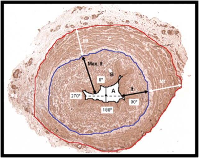
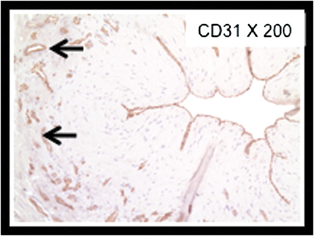
Methods: Vein samples from 12 patients were collected at the time of AV access creation near the site of AV anastomosis. Histological, immunohistochemistry and morphometric studies were performed on these vein samples.

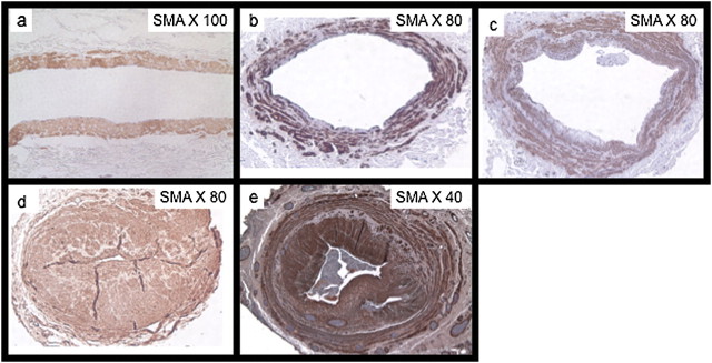
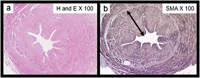
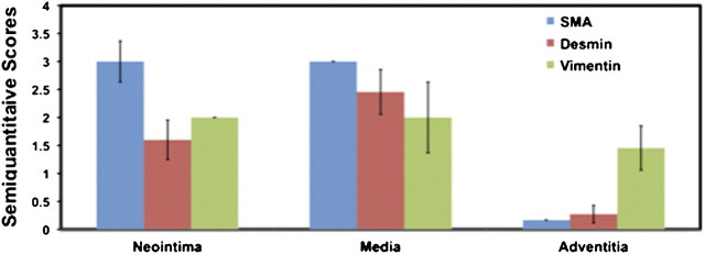
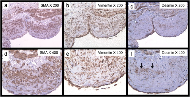
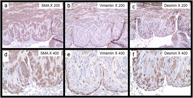
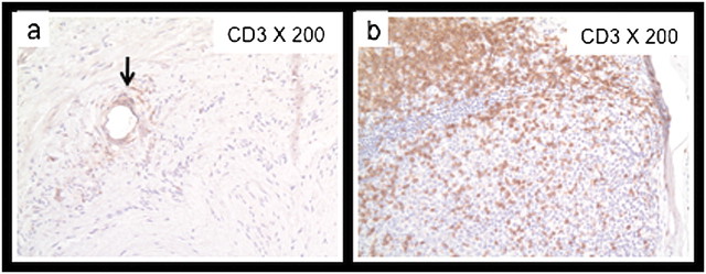
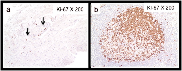
Results: Examination of the tissue specimens obtained at the time of surgery showed neointimal hyperplasia in 10 of 12 specimens, ranging from minimal to very severe. The majority of cells within the neointima were myofibroblasts with a minority of contractile smooth muscle cells present.

Conclusion: Our work represents a detailed description of the morphometric and cellular phenotypic lesions present in the veins of CKD and ESRD patients, prior to dialysis access placement. These studies (i) suggest the future possibility of a new predictive marker (pre-existing venous neointimal hyperplasia) for AV dialysis access dysfunction and (ii) open the door for the future development of novel local therapies for optimization of the venous substrate on which the dialysis access is created.


Figures

Fig. 1
Fig. 1
Morphometric analyses. Methods used for morphometric analyses as described in text. Area enclosed by the solid black line is the luminal area, area enclosed by the blue line is the combined luminal plus neointimal area and the area enclosed by the red is the luminal + intima–media area. Double-headed black arrows show intimal thickness (lt) and maximal intimal thickness (Max It) The white double-headed arrows represent medial thickening measurements for average and maximal intimal to medial thickness (Mt) ratios.
Fig. 2
Fig. 2
Histopathology of vein specimens from normal patients to stenosed AVF. (a) Shows normal human vein in a patient with no CKD. Note the absence of medial thickness and neointimal hyperplasia (bd) shows SMA sections of patients with advanced CKD at the time of AV access placement. Note that neointimal hyperplasia in patients is variable from minimal neointimal hyperplasia to very severe lesions (e) shows a human vein in a patient with ESRD with a stenotic AV fistula. Note the aggressive thickening of the neointima and media and significant luminal stenosis that is similar to the lesion prior to access placement in some patients (d).
Fig. 3
Fig. 3
H and E and SMA of severe pre-existing neointimal hyperplasia. (A) H&E) and (B) SMA on sequential sections of vein specimen obtained at the time of AV access surgery. Note the very severe degree of neointimal hyperplasia [black double-headed arrows in (B)].
Fig. 4
Fig. 4
Cellular phenotyping of venous specimens. Note that the majority of cells in the neointima appear to be SMA-positive, vimentin-positive myofibroblasts, albeit with a reasonable number of SMA-positive, desmin-positive contractile smooth muscle cells. The majority of the cells within the media appear to be SMA-positive, desmin-positive contractile smooth muscle cells with a smaller number of SMA-positive, vimentin-positive myofibroblasts. The adventitia is primarily composed of vimentin-positive, SMA-negative, desmin-negative fibroblasts.
Fig. 5
Fig. 5
Cellular phenotyping of neointimal cells representing predominantly myofibroblasts: the expression of smooth muscle cell actin (a), vimentin (b) and desmin (c) within the sequential sections of a patient with neointimal hyperplasia. The majority of cells seen on high-power view are SMA-positive (d), vimentin-positive (e) myofibroblasts. However, note that a high-power view does show some desmin-positive cells (arrows) (f) within the intima–media thickening.
Fig. 6
Fig. 6
Cellular phenotyping of neointimal hyperplasia representing predominantly contractile smooth muscle cells. The expression of alpha smooth muscle actin (a), vimentin (b) and desmin (c) within the same section of a patient with neointimal hyperplasia. The majority of cells seen on high-power view in this section as opposed to Figure 5 are SMA-positive (d), vimentin-negative (e) and desmin-positive (f) contractile smooth muscle cells.
Fig. 7
Fig. 7
CD3 stain for lymphocytes. (a) Shows minimal staining of a representative vein sample with neointimal hyperplasia for CD3. Note the perivascular pattern of lymphocytic infiltration (arrow). (b) Positive control of human tonsil demonstrating significant staining for CD3.
Fig. 8
Fig. 8
Ki-67 stain for proliferating cells. (a) Shows minimal staining for a representative vein sample with neointimal hyperplasia with only a few proliferating cells present at the intima–media junction. (b) Positive control of human tonsil showing significant staining for proliferating cells.
Fig. 9
Fig. 9
CD31 stain for endothelial cells. Representative vein sample with severe neointimal hyperplasia showing staining within the endothelium for CD31. Note also the ring of microvessels within the adventitia (arrows). We often used these vessels to determine the outer border of the media.

References

    1. Roy-Chaudhury P, Sukhatme VP, Cheung AK. Hemodialysis vascular access dysfunction: a cellular and molecular viewpoint. J Am Soc Nephrol. 2006;17:1112–1127. - PubMed
    1. Lee T, Roy-Chaudhury P. Advances and new frontiers in the pathophysiology of venous neointimal hyperplasia and dialysis access stenosis. Adv Chronic Kidney Dis. 2009;16:329–338. - PMC - PubMed
    1. Roy-Chaudhury P, Lee TC. Vascular stenosis: biology and interventions. Curr Opin Nephrol Hypertens. 2007;16:516–522. - PubMed
    1. Allan JC, Bertram K, Charles H, et al. United States Renal Data System 2006 Annual Data Report Abstract. Am J Kidney Dis. 2006;49:A6–A7. - PubMed
    1. Dember LM, Beck GJ, Allon M, et al. Effect of clopidogrel on early failure of arteriovenous fistulas for hemodialysis: a randomized controlled trial. JAMA. 2008;299:2164–2171. - PMC - PubMed