. 2010 Oct-Dec;6(4):263-9.
doi: 10.4161/org.6.4.12682.

Microdissection of the gene expression codes driving nephrogenesis

Affiliations

Microdissection of the gene expression codes driving nephrogenesis

S Steven Potter et al. Organogenesis. 2010 Oct-Dec.

Abstract

The kidney represents an excellent model system for learning the principles of organogenesis. It is intermediate in complexity, and employs many commonly used developmental processes. As such, kidney development has been the subject of intensive study, using a variety of techniques, including in situ hybridization, organ culture and gene targeting, revealing many critical genes and pathways. Nevertheless, proper organogenesis requires precise patterns of cell type specific differential gene expression, involving very large numbers of genes. This review is focused on the use of global profiling technologies to create an atlas of gene expression codes driving development of different mammalian kidney compartments. Such an atlas allows one to select a gene of interest, and to determine its expression level in each element of the developing kidney, or to select a structure of interest, such as the renal vesicle, and to examine its complete gene expression state. Novel component specific molecular markers are identified, and the changing waves of gene expression that drive nephrogenesis are defined. As the tools continue to improve for the purification of specific cell types and expression profiling of even individual cells it is possible to predict an atlas of gene expression during kidney development that extends to single cell resolution.


Figures
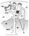
Figure 1
Figure 1
Diagram of kidney development. The capping mesenchyme (CM) is induced by the cortical collecting duct (CCD) to undergo a mesenchyme to epithelia transition and form the renal vesicle (RV), which clefts, grows and fuses with the ureteric bud to form the S-shaped body (S), which grows and segments to form the nephron, with a renal corpuscle (RC), proximal tubule (PT) and loop of Henle (H). Other components include the medullary collecting duct (MCD), medullary interstitium or stroma, (MI) and cortical interstitium (CI). This diagram is reproduced with permission from reference and was derived from reference .
Figure 2
Figure 2
Transcriptional regulatory pathway of genes with elevated expression in the E11.5 metanephric mesenchyme. GeneSpring GX 11.0 was used to select 91 genes with the strongest E11.5 metanephric mesenchyme specific expression and to perform a transcriptional regulatory pathways analysis. Proteins circled in blue are among those with elevated metanephric mesenchyme expression.
Figure 3
Figure 3
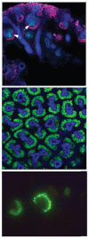
Compartment specific GFP expression in transgenic mice. Top part: Shows restricted GFP expression in the central region of the S-shaped body in Hes5-GFP mice (arrows). For context, red shows Wt1 expression in capping mesenchyme and podocyte progenitors. Blue shows E-cadherin staining of epithelial structures. Middle part: Shows GFP expression restricted to the capping mesenchyme for Crym-GFP mice. Blue marks E-cadherin expression of ureteric buds. Bottom part: Shows podocyte specific expression of GFP in an early glomerulus of a MafB-GFP transgenic mouse.

References

    1. Costantini F, Kopan R. Patterning a complex organ: branching morphogenesis and nephron segmentation in kidney development. Dev Cell. 2010;18:698–712. - PMC - PubMed
    1. Gall JG, Pardue ML. Formation and detection of RNA-DNA hybrid molecules in cytological preparations. Proc Natl Acad Sci USA. 1969;63:378–383. - PMC - PubMed
    1. Jackson DA, Symons RH, Berg P. Biochemical method for inserting new genetic information into DNA of Simian Virus 40: circular SV40 DNA molecules containing lambda phage genes and the galactose operon of Escherichia coli. Proc Natl Acad Sci USA. 1972;69:2904–2909. - PMC - PubMed
    1. Sanger F, Nicklen S, Coulson AR. DNA sequencing with chain-terminating inhibitors. Proc Natl Acad Sci USA. 1977;74:5463–5467. - PMC - PubMed
    1. Gordon JW, Scangos GA, Plotkin DJ, Barbosa JA, Ruddle FH. Genetic transformation of mouse embryos by microinjection of purified DNA. Proc Natl Acad Sci USA. 1980;77:7380–7384. - PMC - PubMed

Publication types