

Renal cell carcinoma with double synchronous contralateral adrenal metastases
- PMID: 21221210
- PMCID: PMC3016436
- DOI: 10.4111/kju.2010.51.12.879
Renal cell carcinoma with double synchronous contralateral adrenal metastases
Abstract
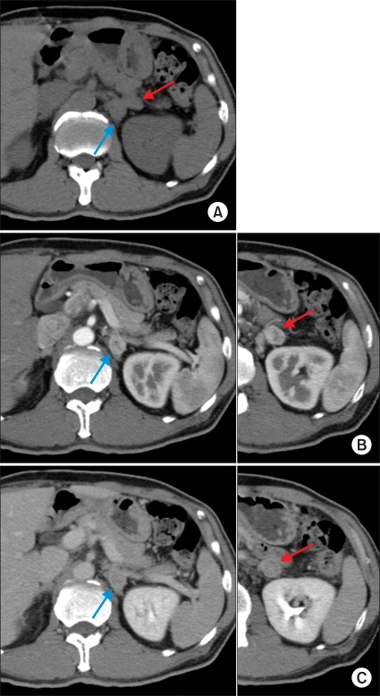
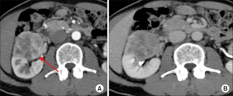
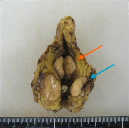
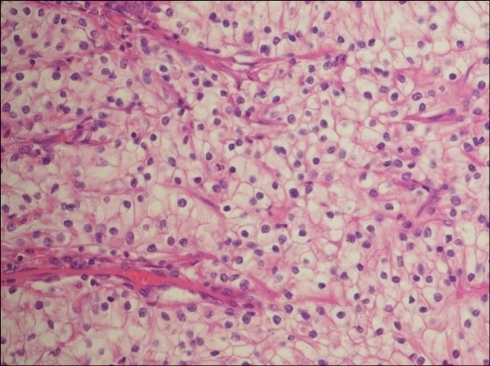
A 63-year-old male patient visited our hospital with a right incidental renal tumor, which was found by ultrasonography for the follow-up study of chronic hepatitis B virus infection and diabetes mellitus. Consecutive computed tomography revealed a right renal tumor and two left adrenal tumors. Further systemic imaging study and hormonal examination suggested one right renal cell carcinoma and left adrenal metastases. We performed right nephrectomy and left adrenalectomy. The pathological diagnoses of all resected tumors were renal cell carcinoma. The patient has been in good health without any recurrence for 12 months since the operation. In patients with renal cell carcinoma, contralateral adrenal metastasis is usually associated with multiple metastases to other organs. There are a few cases of solitary and synchronous contralateral adrenal metastasis in the English literature. To our knowledge, this is the first report of a case of renal cell carcinoma with double synchronous contralateral adrenal metastases.
Keywords: Adrenal glands; Neoplasm metastasis; Neoplasms; Renal cell carcinoma; Synchronous neoplasms.
Conflict of interest statement
The authors have nothing to disclose.
Figures
References
-
- Lau WK, Zinche H, Lohse CM, Cheville JC, Weaver AL, Blute ML. Contralateral adrenal metastasis of renal cell carcinoma: treatment, outcome and a review. BJU Int. 2003;91:775–779. - PubMed
-
- Saitoh H, Nakayama M, Nakamura K, Satoh T. Distant metastasis of renal adenocarcinoma in nephrectomized cases. J Urol. 1982;127:1092–1095. - PubMed
-
- Sapienza P, Stipa F, Lucandri G, Baratti L, Delfino M, Mingazzini PL. Renal carcinoma with a solitary synchronous contralateral adrenal metastasis: a case report. Anticancer Res. 1997;17:743–747. - PubMed
-
- Utsumi T, Suzuki H, Nakamura K, Kim W, Kamijima S, Awa Y, et al. Renal cell carcinoma with a huge solitary metastasis to the contralateral adrenal gland: a case report. Int J Urol. 2008;15:1077–1079. - PubMed
-
- Abi-add AS, Belldegrum A, Dekernion JB. Renal cell carcinoma. Physiology, diagnosis, and therapy. World J Urol. 1991;9:168–172.
LinkOut - more resources
Full Text Sources
