

Frontotemporal dementia caused by CHMP2B mutations
- PMID: 21222599
- PMCID: PMC3182073
- DOI: 10.2174/156720511795563764
Frontotemporal dementia caused by CHMP2B mutations
Abstract
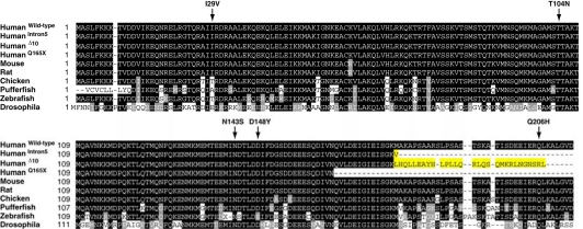
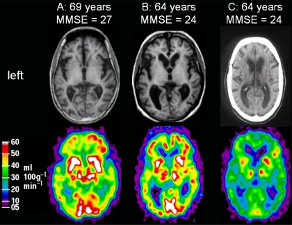
CHMP2B mutations are a rare cause of autosomal dominant frontotemporal dementia (FTD). The best studied example is frontotemporal dementia linked to chromosome 3 (FTD-3) which occurs in a large Danish family, with a further CHMP2B mutation identified in an unrelated Belgian familial FTD patient. These mutations lead to C-terminal truncations of the CHMP2B protein and we will review recent advances in our understanding of the molecular effects of these mutant truncated proteins on vesicular fusion events within the endosome-lysosome and autophagy degradation pathways. We will also review the clinical features of FTD caused by CHMP2B truncation mutations as well as new brain imaging and neuropathological findings. Finally, we collate the current data on CHMP2B missense mutations, which have been reported in FTD and motor neuron disease.
Figures
References
-
- Neary D, Snowden JS, Gustafson L, Passant U, Stuss D, Black S, et al. Frontotemporal lobar degeneration: a consensus on clinical diagnostic criteria. Neurology. 1998;51:1546–1554. - PubMed
-
- McKhann GM, Albert MS, Grossman M, Miller B, Dickson D, Trojanowski JQ. Clinical and pathological diagnosis of frontotemporal dementia: report of the Work Group on Frontotemporal Dementia and Pick's Disease. Arch Neurol. 2001;58:1803–1809. - PubMed
-
- Lillo P, Hodges JR. Frontotemporal dementia and motor neurone disease: overlapping clinic-pathological disorders. J Clin Neurosci. 2009;16:1131–1135. - PubMed
-
- Josephs KA, Petersen RC, Knopman DS, Boeve BF, Whitwell JL, Duffy JR, et al. Clinicopathologic analysis of frontotemporal and corticobasal degenerations and PSP. Neurology. 2006;66:41–48. - PubMed
Publication types
MeSH terms
Substances
Grants and funding
LinkOut - more resources
Full Text Sources