. 2011 Jan 11;13(1):R3.
doi: 10.1186/ar3221.

Toll-like receptor 3 upregulation by type I interferon in healthy and scleroderma dermal fibroblasts

Affiliations

Toll-like receptor 3 upregulation by type I interferon in healthy and scleroderma dermal fibroblasts

Sandeep K Agarwal et al. Arthritis Res Ther. .

Abstract

Introduction: Increased levels of genes in the type I interferon (IFN) pathway have been observed in patients with systemic sclerosis (SSc), or scleroderma. How type I IFN regulates the dermal fibroblast and its participation in the development of dermal fibrosis is not known. We hypothesized that one mechanism by which type I IFN may contribute to dermal fibrosis is through upregulation of specific Toll-like receptors (TLRs) on dermal fibroblasts. Therefore, we investigated the regulation of TLR expression on dermal fibroblasts by IFN.

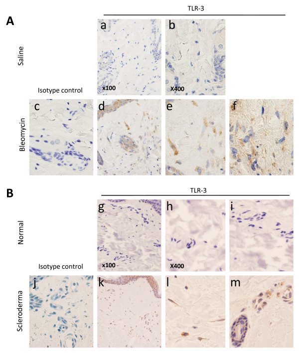
Methods: The expression of TLRs was assessed in cultured dermal fibroblasts from control and SSc patients stimulated with IFNα2. The ability of IFNα2 to regulate TLR-induced interleukin (IL)-6 and CC chemokine ligand 2 production was also assessed. Immunohistochemical analyses were performed to determine whether TLR3 was expressed in skin biopsies in the bleomycin-induced skin fibrosis model and in patients with SSc.

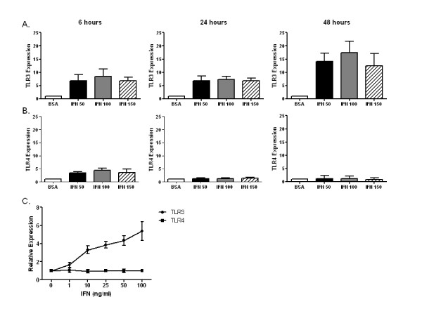
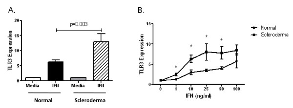
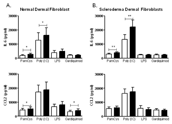
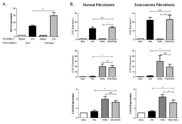
Results: IFNα2 increased TLR3 expression on human dermal fibroblasts, which resulted in enhanced TLR3-induced IL-6 production. SSc fibroblasts have an augmented TLR3 response to IFNα2 relative to control fibroblasts. Pretreatment of fibroblasts with transforming growth factor (TGF)-β increased TLR3 induction by IFNα2, but coincubation of TGF-β did not alter TLR3 induction by IFN. Furthermore, IFNα2 inhibits but does not completely block the induction of connective tissue growth factor and collagen expression by TGF-βin fibroblasts. TLR3 expression was observed in dermal fibroblasts and inflammatory cells from skin biopsies from patients with SSc as well as in the bleomycin-induced skin fibrosis model.

Conclusions: Type I IFNs can increase the inflammatory potential of dermal fibroblasts through the upregulation of TLR3.


Figures

Figure 1
Figure 1
Toll-like receptor 3 (TLR3) upregulation by interferon α (IFNα). Dermal fibroblasts from healthy control skin were cultured in vitro with IFNα (50-150 ng/mL) or 0.1% bovine serum albumin (BSA) for 6, 24 and 48 hours. Total RNA was harvested, and (A) TLR3 and (B) TLR4 mRNA levels were determined by performing quantitative real-time polymerase chain reaction (qRT-PCR) assays. IFN induced TLR3 upregulation at 6, 24 and 48 hours. TLR4 upregulation was noted only at 6 hours. (C) Dose-response curve for TLR3 upregulation by IFNα (0-100 ng/mL) for 24 hours in healthy control dermal fibroblasts. n = 3 control cell lines.
Figure 2
Figure 2
Comparison of TLR3 upregulation by IFNα in healthy control and systemic sclerosis (SSc), or scleroderma, dermal fibroblasts. (A) Dermal fibroblasts were stimulated for 24 hours with 50 ng/mL IFNα, and TLR3 was determined by performing qRT-PCR assays. The magnitude of induction of TLR3 expression by IFNα was significantly greater in dermal fibroblasts from patients with SSc (n = 11) than in those from healthy controls (n = 25; P = 0.003). (B) SSc dermal fibroblasts have a greater magnitude of upregulation of TLR3 with IFN at concentrations ranging from 1 to 100 ng/mL (n = 4 in each group; *P < 0.05 (Wilcoxon signed-rank test)).
Figure 3
Figure 3
IFN increases TLR3-induced interleukin (IL)-6 production in cultured dermal fibroblasts. (A) Healthy control fibroblasts (n = 10) and (B) SSc dermal fibroblasts (n = 10) were preincubated with media alone or with 50 ng/mL IFNα for 24 hours, washed and then stimulated with Pam3CysK4 (TLR2 agonist); polyinosinic:polycytidylic acid, or poly(I:C) (TLR3 agonist); lipopolysaccharide (TLR4 agonist) and Gardiquimod (TLR7/8 agonist; [1-(4-amino-2-ethylaminomethylimidazo[4,5-c]quinolin-1-yl)-2-methylpropan-2-ol]) for 48 hours (10 μg/mL). Culture supernatants were assessed for IL-6 and CC chemokine ligand 2 (CCL2). Preincubation with IFNα increased poly(I:C)-stimulated IL-6 but not CCL2 production from healthy control and SSc dermal fibroblasts. *P < 0.05, **P < 0.01 (Wilcoxon signed-rank test).
Figure 4
Figure 4
Cross-regulation of IFNα and TGFβ in dermal fibroblasts. (A) Healthy control dermal fibroblasts were cultured in 10 ng/mL TGFβ for 72 hours to induce myofibroblast differentiation in vitro. After 72 hours, cultures were washed and subsequently stimulated with 50 ng/mL IFN for 24 hours. Total RNA was analyzed for TLR3 by qRT-PCR assay. Preincubation with TGFβ resulted in a greater induction of TLR3 by IFN compared with fibroblasts preincubated in 0.1% BSA (n = 7; P = 0.02). (B) Dermal fibroblasts were incubated with 50 ng/mL IFN, 10 ng/mL TGF-β or both cytokines for 24 hours. Total RNA was analyzed for TLR3, connective tissue growth factor (CTGF), and collagen type I, α1 (COL1A1) expression by qRT-PCR assay. Coincubation of fibroblasts with IFN and TGF-β did not alter the expression of TLR3 compared with IFN alone. IFN did not alter TGF-β-induced CTGF expression but did slightly reduce COL1A1 expression in SSc dermal fibroblasts. n = 7, *P < 0.05, n.s. = not significant (Wilcoxon signed-rank test).
Figure 5
Figure 5
Immunohistochemical analyses of TLR3 expression in dermal fibrosis. Immunohistochemical analyses were performed using rabbit polyclonal antibodies against TLR3 (histograms a, b, d-f, g-i, k-m) or isotype control (histograms c and j). (A) Skin biopsies from mice injected with bleomycin, but not saline, demonstrated expression of TLR3 in the dermis (panel d), which localized to fibroblast-like cells (histogram e) and inflammatory cells (histogram f). n = 3 saline, n = 3 bleomycin. (B) Skin biopsies from control skin (n = 4) and SSc skin (n = 4) demonstrated TLR3 expression in the dermis of SSc skin (histogram k), which localized to fibroblast-like cells and inflammatory cells (histogram l) as well as to endothelial cells (histogram m) in SSc but no control skin.

References

    1. Varga J, Abraham D. Systemic sclerosis: a prototypic multisystem fibrotic disorder. J Clin Invest. 2007;117:557–567. doi: 10.1172/JCI31139. - DOI - PMC - PubMed
    1. Varga J. Systemic sclerosis: an update. Bull NYU Hosp Jt Dis. 2008;66:198–202. - PubMed
    1. Kissin EY, Merkel PA, Lafyatis R. Myofibroblasts and hyalinized collagen as markers of skin disease in systemic sclerosis. Arthritis Rheum. 2006;54:3655–3660. doi: 10.1002/art.22186. - DOI - PubMed
    1. Tomasek JJ, Gabbiani G, Hinz B, Chaponnier C, Brown RA. Myofibroblasts and mechano-regulation of connective tissue remodelling. Nat Rev Mol Cell Biol. 2002;3:349–363. doi: 10.1038/nrm809. - DOI - PubMed
    1. Feghali CA, Bost KL, Boulware DW, Levy LS. Control of IL-6 expression and response in fibroblasts from patients with systemic sclerosis. Autoimmunity. 1994;17:309–318. doi: 10.3109/08916939409010671. - DOI - PubMed

Publication types

MeSH terms