. 2011 Jan 12:8:11.
doi: 10.1186/1743-422X-8-11.

Full-length Ebola glycoprotein accumulates in the endoplasmic reticulum

Affiliations

Full-length Ebola glycoprotein accumulates in the endoplasmic reticulum

Suchita Bhattacharyya et al. Virol J. .

Abstract

The Filoviridae family comprises of Ebola and Marburg viruses, which are known to cause lethal hemorrhagic fever. However, there is no effective anti-viral therapy or licensed vaccines currently available for these human pathogens. The envelope glycoprotein (GP) of Ebola virus, which mediates entry into target cells, is cytotoxic and this effect maps to a highly glycosylated mucin-like region in the surface subunit of GP (GP1). However, the mechanism underlying this cytotoxic property of GP is unknown. To gain insight into the basis of this GP-induced cytotoxicity, HEK293T cells were transiently transfected with full-length and mucin-deleted (Δmucin) Ebola GP plasmids and GP localization was examined relative to the nucleus, endoplasmic reticulum (ER), Golgi, early and late endosomes using deconvolution fluorescent microscopy. Full-length Ebola GP was observed to accumulate in the ER. In contrast, GPΔmucin was uniformly expressed throughout the cell and did not localize in the ER. The Ebola major matrix protein VP40 was also co-expressed with GP to investigate its influence on GP localization. GP and VP40 co-expression did not alter GP localization to the ER. Also, when VP40 was co-expressed with the nucleoprotein (NP), it localized to the plasma membrane while NP accumulated in distinct cytoplasmic structures lined with vimentin. These latter structures are consistent with aggresomes and may serve as assembly sites for filoviral nucleocapsids. Collectively, these data suggest that full-length GP, but not GPΔmucin, accumulates in the ER in close proximity to the nuclear membrane, which may underscore its cytotoxic property.


Figures

Figure 1
Figure 1
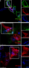
Full-length Ebola GP localizes in the ER in close proximity to the nuclear membrane. A. HEK293T cells were transiently transfected with 10 μg GP (pCB6-EbGP) plasmid for 24 hours. Cells were fixed and stained for DNA (blue) using Hoechst, nuclear pore complex (NPC, red) proteins using mouse monoclonal antibody 414 (Covance Research Products), and GP (green) using the neutralizing human monoclonal antibody (KZ52) labeled with a Zenon labeling kit (Molecular Probes). Scale bar represents 15 μm. Side panels show individual fluorescent channels from the boxed region in the image. B. HEK293T cells were transiently transfected for 24 hours with 8 μg GP and 2 μg pDsRed2-ER vector (Clontech) that labels the endoplasmic reticulum (ER, red). Cells were fixed and stained for late endosomes using a mouse monoclonal antibody targeting CD63 (BD Biosciences, blue), in accordance with a previous publication [43]. GP was stained as above (green). Scale bar represents 15 μm. Side panels show individual fluorescent channels from the boxed region in the image.
Figure 2
Figure 2
Ebola GP lacking the mucin-like region does not accumulate in the ER. A. HEK293T cells were transiently transfected for 24 hours with 8 μg GPΔmucin (pCDNA6-EbGPΔmucin-mutΔ1234) and 2 μg pDsRed2-ER vector (Clontech) that labels the endoplasmic reticulum (ER, red). Cells were fixed and stained for NPC proteins (blue) using mouse monoclonal antibody 414 (Covance Research Products) and GPΔmucin (green) using the KZ52 neutralizing human monoclonal antibody labeled with a Zenon labeling kit (Molecular Probes). Scale bar represents 15 μm. Side panels show individual fluorescent channels from the boxed region in the image. B. HEK293T cells were transiently transfected for 24 hours with 8 μg GPΔmucin and 2 μg pEYFP-Golgi vector (Clontech) that labels the trans-medial region of the Golgi apparatus (blue). Cells were fixed and stained for early endosomes using a mouse monoclonal antibody against EEA1 (BD Biosciences, green) and for GPΔmucin as above (red). Scale bar represents 15 μm. Side panels show individual fluorescent channels from the boxed region in the image. C. HEK293T cells were transiently transfected with 10 μg GPΔmucin for 24 hours. Cells were fixed and stained for late endosomes using a mouse monoclonal antibody targeting CD63 (BD Biosciences, blue) and for GPΔmucin as above (red). Scale bar represents 15 μm. Side panels show individual fluorescent channels from the boxed region in the image.
Figure 3
Figure 3
Co-expression of Ebola GP, VP40 and NP in HEK293T cells does not alter localization of individual proteins. A. HEK293T cells were co-transfected with 5 μg Ebola GP and 5 μg eGFP-VP40 plasmids for 24 hours. Cells were fixed and stained for NPC proteins using mouse monoclonal antibody 414 (Covance Research Products, blue) and GP (red) using KZ52 neutralizing human monoclonal antibody labeled with a Zenon labeling kit (Molecular Probes). Green represents VP40. Scale bar represents 15 μm. Upper and lower side panels show VP40 and GP fluorescence respectively, from the boxed region in the image. B. HEK293T cells were transfected with 10 μg Ebola NP plasmid for 24 hours. Cells were fixed and stained for DNA using Hoechst (blue), NP using a mouse monoclonal antibody targeting NP (red) and vimentin using a mouse monoclonal antibody targeting vimentin (green). Scale bar represents 5 μm. Upper and lower side panels show the vimentin and DNA fluorescence and the NP and DNA fluorescence respectively, from the boxed region in the image. C. HEK293T cells were co-transfected with 5 μg Ebola NP and 5 μg eGFP-VP40 plasmids for 24 hours. Cells were fixed and stained for NP using a mouse monoclonal antibody targeting NP (red) and DNA using Hoechst (blue). Green represents VP40. Scale bar represents 5 μm. Upper and lower side panels show the VP40 and DNA fluorescence and the NP and DNA fluorescence respectively. D. HEK293T cells were co-transfected with 5 μg Ebola NP and 5 μg eGFP-VP40 plasmids for 24 hours. Cells were fixed and stained for NP using a mouse monoclonal antibody targeting NP (blue) and filamentous actin using Texas Red Phalloidin (Invitrogen, red). Green represents VP40. Scale bar represents 15 μm. Side panels show the individual fluorescent channels from the boxed region in the image.

References

    1. Takada A, Robison C, Goto H, Sanchez A, Murti KG, Whitt MA, Kawaoka Y. A system for functional analysis of Ebola virus glycoprotein. Proc Natl Acad Sci USA. 1997;94:14764–14769. doi: 10.1073/pnas.94.26.14764. - DOI - PMC - PubMed
    1. Wool-Lewis RJ, Bates P. Characterization of Ebola virus entry by using pseudotyped viruses: identification of receptor-deficient cell lines. J Virol. 1998;72:3155–3160. - PMC - PubMed
    1. Chan SY, Ma MC, Goldsmith MA. Differential induction of cellular detachment by envelope glycoproteins of Marburg and Ebola (Zaire) viruses. J Gen Virol. 2000;81:2155–2159. - PubMed
    1. Simmons G, Wool-Lewis RJ, Baribaud F, Netter RC, Bates P. Ebola virus glycoproteins induce global surface protein down-modulation and loss of cell adherence. J Virol. 2002;76:2518–2528. doi: 10.1128/jvi.76.5.2518-2528.2002. - DOI - PMC - PubMed
    1. Takada A, Watanabe S, Ito H, Okazaki K, Kida H, Kawaoka Y. Downregulation of beta1 integrins by Ebola virus glycoprotein: implication for virus entry. Virology. 2000;278:20–26. doi: 10.1006/viro.2000.0601. - DOI - PubMed

Publication types

LinkOut - more resources