. 2011 Jan;178(1):128-39.
doi: 10.1016/j.ajpath.2010.11.011. Epub 2010 Dec 23.

Cytoglobin, a novel member of the globin family, protects kidney fibroblasts against oxidative stress under ischemic conditions

Affiliations

Cytoglobin, a novel member of the globin family, protects kidney fibroblasts against oxidative stress under ischemic conditions

Hiroshi Nishi et al. Am J Pathol. 2011 Jan.

Abstract

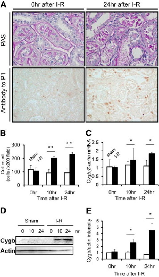
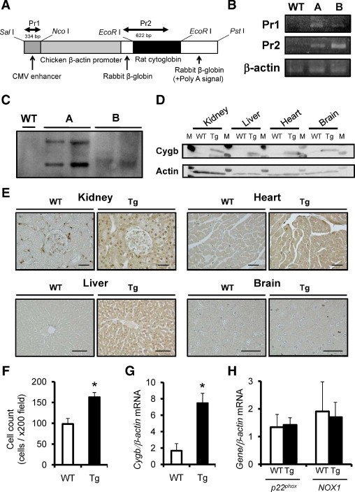
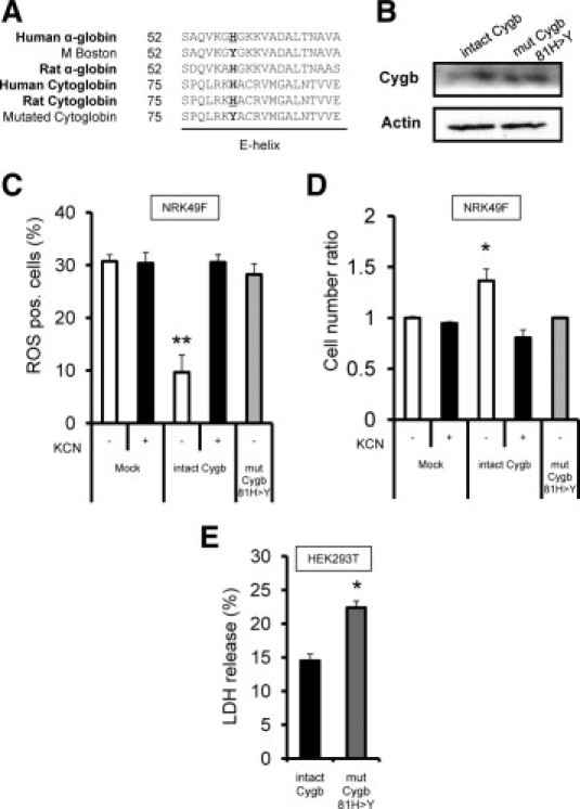
Cytoglobin (Cygb) is a novel member of the vertebrate globin superfamily. Although it is expressed in splanchnic fibroblasts of various organs, details of its function remain unknown. In the present study, kidney ischemia-reperfusion (I/R) increased the number of Cygb-positive cells per area and up-regulated Cygb mRNA and protein expression in kidney cortex tissues. Similarly, hypoxia up-regulated Cygb expression in cultured rat kidney fibroblasts. The biological function of Cygb in vivo was evaluated in Cygb-overexpressing transgenic rats. Renal dysfunction and histologic damage after renal I/R were ameliorated (mean [SE] serum urea nitrogen concentration after I/R injury, 260.6 [44.9] mg/dL in wild-type rats versus 101.0 [36.0] mg/dL in transgenic rats; P < 0.05) in association with improvement of oxidative stress. Primary cultured fibroblasts from Cygb transgenic rat kidney were resistant to exogenous oxidant stimuli, and treatment of immortalized kidney fibroblasts with Cygb-small interfering RNA (siRNA) enhanced cellular oxidant stress and subsequently decreased cell viability (cell count ratio after exposure to hydrogen peroxide, 35.9% [1.6%] in control-siRNA-treated cells versus 25.5% [2.0%] in Cygb-siRNA-treated cells; P < 0.05). Further, chemical or mutant disruption of heme in Cygb impaired its antioxidant properties, which suggests that the heme of Cygb per se possesses a radical scavenging function. These findings show for the first time, to our knowledge, that Cygb serves as a defensive mechanism against oxidative stress both in vitro and in vivo.


Figures

Figure 1
Figure 1
Generation and characterization of rabbit polyclonal antibodies against rat Cygb polypeptides. A: Rat Cygb consists of 190 amino acids with a molecular weight of 21,496. Two different antibodies to the corresponding amino acid sequences (full line, P1; dashed line, P2) were used. B: Titration of the polyclonal antibody P1. ELISA of the polyclonal antibody revealed an increase in light absorbance in a dose-dependent manner (full line, closed circle). Isotype IgG control (dashed line, open circle). C: Immunoblot analysis of rat kidney cortex using the antibodies specifically identified Cygb protein.
Figure 2
Figure 2
Immunostaining analysis of Cygb in the normal rat kidney. A: Immunostaining of Cygb with anti-P1 antibody was observed in interstitial fibroblasts of normal rat kidney. Scale bar = 50 μm. Inset, High-power magnification showed diffuse distribution of Cygb in the cell body. Double scale bar = 10 μm. B: Immunostaining of Cygb with anti-P2 antibody was also observed in interstitial fibroblasts and in the mesangial area of normal rat kidney. C: Staining with anti-P1 antibody after antigen retrieval resulted in positive staining in the mesangial area of normal rat kidney. D: Staining for anti-P1 antibody preabsorbed by P1 was negative, confirming its specificity. Scale bar = 50 μm. Images of double immunofluorescence staining with Cygb and cell-specific markers are presented. E: JG12 (vascular endothelial cell) in red and Cygb in green are shown in normal kidney. F: ED1 (macrophage or monocyte) in red, Hoechst in blue, and Cygb in green are shown in the kidney at 48 hours after I/R injury. Arrowheads, macrophages and monocytes. Original magnification of E and F ×200.
Figure 3
Figure 3
Cygb expression in rat kidney cortex after I/R injury. A: PAS staining demonstrated that I/R injury induced severe tubular dilation, tubular epithelial injury, debris accumulation, and cast formation at 24 hours after injury (original magnification ×400). Immunohistochemical analysis using anti-P1 showed up-regulation of Cygb expression in the interstitium at the time-point. (original magnification ×200). B: Quantification of Cygb-positive cells in rat kidney tissue showed temporal increases in Cygb staining after I/R (closed column) in comparison with that after sham operation (open column). C: Cygb mRNA expression level was increased at 10 and 24 hours after I/R injury in a time-dependent manner (closed column), whereas no significant change was observed in sham-operated rats (open column). D: Representative immunoblot of Cygb in kidney cortex tissue after I/R. E: Densitometric analysis of immunoblot demonstrated time-dependent up-regulation of Cygb after I/R injury (closed column) but not after sham operation (open column). *P < 0.05/ **P < 0.01.
Figure 4
Figure 4
Establishment and characterization of Cygb-transgenic rats (Tg). A: Cygb transgene construct. Full-length rat Cygb cDNA was subcloned in a vector controlled by the rabbit β-globin gene, including a part of the second intron, the third exon, and the 3′ untranslated region. The positions of primers for PCR analysis are indicated above the construct. B: Identification of rat Cygb transgene by PCR of genomic DNA (line A or B) using primers 1 and 2 (Pr1 and Pr2). C: Southern blot analysis after EcoRI digestion of genomic DNA using probe against transgene sequence. Random integration is demonstrated as distinct position of the bands noted only in mutant lines. D: Overexpression of rat Cygb protein in multiple organs including the kidney, liver, heart, and brain was confirmed using immunoblot analysis. M, molecular weight marker. E: Immunohistochemical analysis also showed enhancement of Cygb protein distribution in these organs of transgenic rats. Scale bar = 50 μm. F: The number of Cygb-positive interstitial cells in the kidney was increased in Cygb-transgenic rats (closed column) compared with wild-type littermates (WT) (open column). G: Overexpression of Cygb mRNA was confirmed in peripheral blood leukocytes of Cygb-transgenic rats (closed column) compared with wild-type littermates (open column). H: Expression of p22phox and NOX1, principal NADPH oxidase components, was examined in the kidney of wild-type rats (open column) and Cygb-transgenic rats (closed column). Transgenic overexpression of Cygb did not affect expression of these enzyme mRNAs. *P < 0.01.
Figure 5
Figure 5
Attenuation of kidney I/R injury by transgenic overexpression of Cygb in rats. Evaluation of physiologic parameters, serum urea nitrogen (A) and serum creatinine concentration (B), demonstrated that renal dysfunction was milder in Cygb-transgenic rats (Tg) (closed column) at 48 hours after kidney I/R injury compared with that in wild-type rats (WT) (open column). C: Histologic analyses of PAS staining also showed improvement in tissue injury at 48 hours after I/R in Cygb-transgenic rats, including improvements in tubular dilation, epithelial detachment, cast formation, and interstitial cell infiltration. D: Inhibition of histologic damage 48 hours after I/R in transgenic rats (closed column) compared with wild-type littermates (open column) was confirmed using a tubulointerstitial injury scoring system. Confirming validity, tubulointerstitial injury scores showed a significant positive correlation with serum urea nitrogen (E) and serum creatinine (F) concentrations in these rats. Scattergram shows results for transgenic (closed circles) and wild-type (open circles) rats. Original magnification ×400. *P < 0.05.
Figure 6
Figure 6
Oxidative stress and attenuation of kidney I/R injury by transgenic overexpression of Cygb in rats. A: Suppressed deposition of oxidative stress markers 4-HNE in kidney tubulointerstitial area was also observed in Cygb-transgenic rats (Tg). Original magnification ×400. Amelioration of deposition of 4-HNE (B) and nitrotyrosine (C) in transgenic rats (closed column) compared with wild-type rats (WT) (open column) was confirmed at morphometric analysis. D: There were only a few ED1-positive cells before I/R injury in both wild-type and transgenic rats. Although I/R injury increased infiltrating cells, the number of ED1-positive cells was fewer in the Cygb-transgenic rats. Original magnification ×200. E: Quantitative analysis showed the reduced number of ED1-positive cell counts in the Cygb-transgenic rats (closed column) compared with wild-type rats (open column). F: PCNA-positive cell counts were evaluated. At baseline, there were few PCNA-positive cells in wild-type (open column) and Cygb-transgenic (closed column) rat kidney. I/R injury induced proliferation of kidney cells, and the number of PCNA-positive cells was less in Cygb-transgenic rats compared with wild-type rats. *P < 0.05. **P < 0.01.
Figure 7
Figure 7
Oxidative stress and overexpression or knockdown of Cygb expression in rat kidney fibroblasts. A: Immunocytochemical analysis showed vimentin expression in primary cultured fibroblasts from rat kidney. Original magnification ×1000. Expression of Cygb was higher in primary cultures of kidney fibroblasts from Cygb-transgenic rats than from wild-type rats, as estimated by immunocytochemical (B) and immunoblot (C) analyses. D: Cell damage under 24-hour exposure with 100 μmol/L of hydrogen peroxide as oxidant stress inducer was evaluated using the lactic dehydrogenase assay. The enzyme release ratio was suppressed in Cygb-transgenic rat kidney fibroblasts (closed column) compared with wild-type fibroblasts (open column). E: During the time course, Cygb-transgenic rat kidney fibroblasts (full line) showed better survival than did wild-type fibroblasts (dashed line) under exposure of 50 μmol/L of hydrogen peroxide, as demonstrated using the MTS assay. F: Cygb-siRNA decreased Cygb protein expression at 24 hours after transfection in NRK49F cells. G: Flow cytometric analysis to detect radical fluorescence in rat kidney fibroblasts treated with hydrogen peroxide showed that hydrogen peroxide increased cellular ROS production in Cygb-siRNA-treated fibroblasts. The ROS-positive cell number in the Cygb-siRNA-treated fibroblasts (closed column) was larger than that in the control-siRNA-treated fibroblasts (open column). H: Viability of cultured fibroblasts treated with Cygb-siRNA when exposed to 300 μmol/L of hydrogen peroxide for 24 hours as evaluated. NRK49F cells treated with Cygb-siRNA (closed column) were more susceptible to extracellular oxidative stimuli than those treated with control siRNA (open column). NS, no significant difference. *P < 0.05.
Figure 8
Figure 8
Decrease in protective properties of Cygb chemical or genetic disruption of heme protein in Cygb. A: For mutagenesis of rat Cygb molecule unable to bind ligands, gene mutation of the hemoglobin α-subunit responsible for human methemoglobinemia was used. A common mutated designation, also called M Boston, naturally occurs as 58His>Tyr in the E helix of human α-globin protein, which corresponds to 81His>Tyr in rat Cygb. B: Plasmid vector expressing intact form of Cygb protein or 58His>Tyr mutated Cygb protein was transfected into NRK49F cells. Immunoblotting analysis with anti-P1 antibody showed equal levels of expression of intact and mutant forms of Cygb for reasonable comparison in transient transfection studies. C: While fibroblasts expressing an intact form of rat Cygb demonstrated lower levels of intracellular ROS under exposure to hydrogen peroxide in comparison with fibroblasts transfected with control vectors (open column), fibroblasts expressing mutated rat Cygb failed to reduce intracellular ROS levels (shaded column). Potassium cyanide added as heme poison to the culture media inhibited ROS reduction in cells expressing intact Cygb but did not affect ROS levels in control cells (closed column). D: The MTS cell counting method demonstrated that the cell viability of intact Cygb-transfected fibroblasts after 6-hour exposure to 1 mmol/L of hydrogen peroxide (open column) was greater than that of mutated Cygb-transfected fibroblasts (shaded column) or of fibroblasts cultured with potassium cyanide (closed column). E: Antioxidant property of Cygb was confirmed in HEK293T cells. The lactic dehydrogenase release assays demonstrated that HEK293T cells expressing intact Cygb protein (closed column) were resistant to 24-hour exposure to 50 μmol/L of hydrogen peroxide compared with cells expressing mutated Cygb (shaded column). *P < 0.05. **P < 0.01.

References

    1. Hardison R.C. A brief history of hemoglobins: plant, animal, protist, and bacteria. Proc Natl Acad Sci USA. 1996;93:5675–5679. - PMC - PubMed
    1. Wittenberg B.A., Wittenberg J.B. Transport of oxygen in muscle. Annu Rev Physiol. 1989;51:857–878. - PubMed
    1. Kawada N., Kristensen D.B., Asahina K., Nakatani K., Minamiyama Y., Seki S., Yoshizato K. Characterization of a stellate cell activation-associated protein (STAP) with peroxidase activity found in rat hepatic stellate cells. J Biol Chem. 2001;276:25318–25323. - PubMed
    1. Trent J.T., III, Hargrove M.S. A ubiquitously expressed human hexacoordinate hemoglobin. J Biol Chem. 2002;277:19538–19545. - PubMed
    1. Burmester T., Ebner B., Weich B., Hankeln T. Cytoglobin: a novel globin type ubiquitously expressed in vertebrate tissues. Mol Biol Evol. 2002;19:416–421. - PubMed

Publication types

MeSH terms