. 2011 Jan;178(1):175-86.
doi: 10.1016/j.ajpath.2010.11.026. Epub 2010 Dec 23.

Bile acids induce inflammatory genes in hepatocytes: a novel mechanism of inflammation during obstructive cholestasis

Affiliations

Bile acids induce inflammatory genes in hepatocytes: a novel mechanism of inflammation during obstructive cholestasis

Katryn Allen et al. Am J Pathol. 2011 Jan.

Abstract

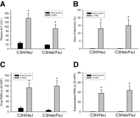
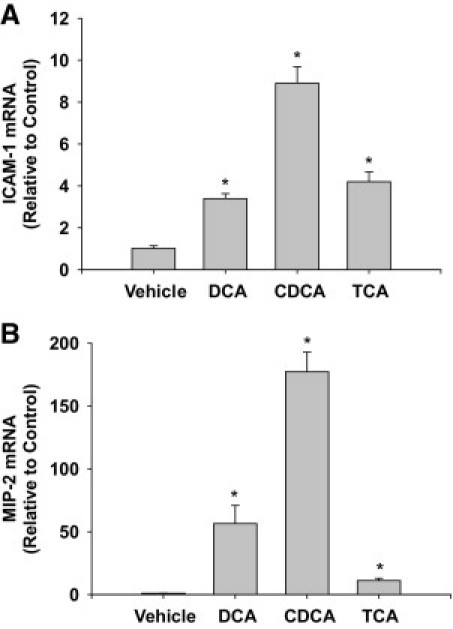
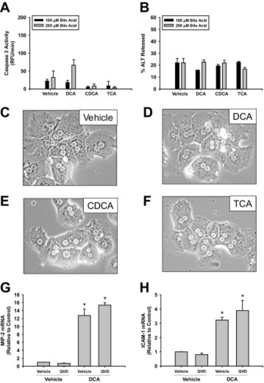
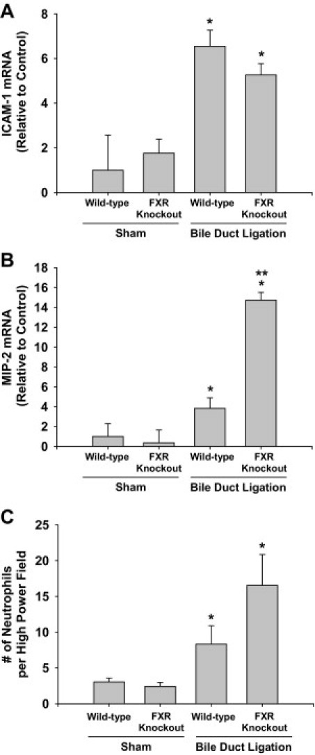
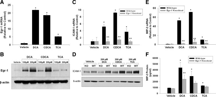
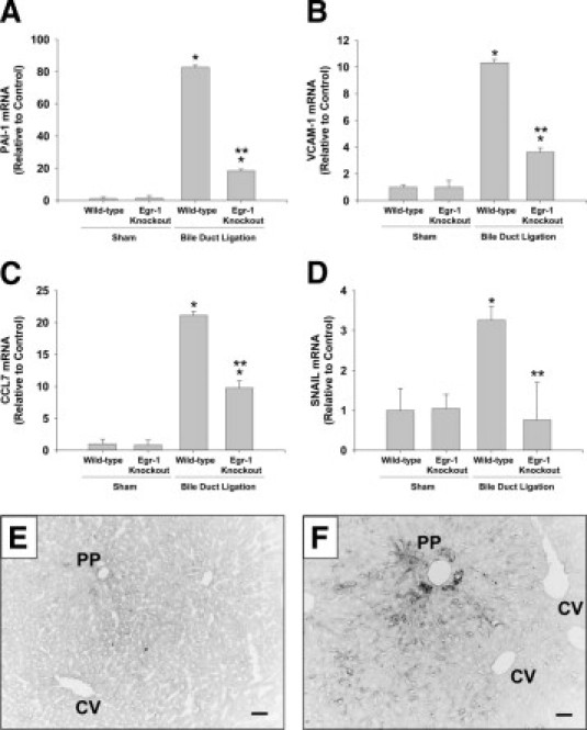
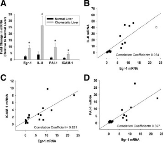
Inflammation contributes to liver injury during cholestasis. The mechanism by which cholestasis initiates an inflammatory response in the liver, however, is not known. Two hypotheses were investigated in the present studies. First, activation of Toll-like receptor 4 (TLR4), either by bacterial lipopolysaccharide or by damage-associated molecular pattern molecules released from dead hepatocytes, triggers an inflammatory response. Second, bile acids act as inflammagens, and directly activate signaling pathways in hepatocytes that stimulate production of proinflammatory mediators. Liver inflammation was not affected in lipopolysaccharide-resistant C3H/HeJ mice after bile duct ligation, indicating that Toll-like receptor 4 is not required for initiation of inflammation. Treatment of hepatocytes with bile acids did not directly cause cell toxicity but increased the expression of numerous proinflammatory mediators, including cytokines, chemokines, adhesion molecules, and other proteins that influence immune cell levels and function. Up-regulation of several of these genes in hepatocytes and in the liver after bile duct ligation required early growth response factor-1, but not farnesoid X receptor. In addition, early growth response factor-1 was up-regulated in the livers of patients with cholestasis and correlated with levels of inflammatory mediators. These data demonstrate that Toll-like receptor 4 is not required for the initiation of acute inflammation during cholestasis. In contrast, bile acids directly activate a signaling network in hepatocytes that promotes hepatic inflammation during cholestasis.


Figures

Figure 1
Figure 1
Role of Toll-like receptor 4 (TLR4) in inflammation in the liver during cholestasis. C3H/HeJ (TLR4 mutant) and C3Heb/FeJ (TLR4 wild-type) mice were subjected to bile duct ligation (BDL) or sham operation. Three days later, plasma alanine aminotransferase (ALT) (A), % area of necrosis (B), neutrophil (PMN) accumulation (C), and PMN extravasation (D) were measured. Data are expressed as mean ± SEM; n = .6. Values significantly different (*P < 0.05) from sham-operated mice. HPF, high-power field.
Figure 2
Figure 2
Up-regulation of intercellular adhesion molecule-1 (ICAM-1) and macrophage inflammatory protein-2 (MIP-2) in bile acid-treated hepatocytes. Hepatocytes were isolated from mice and treated with 200 μmol/L deoxycholic acid (DCA), chenodeoxycholic acid (CDCA), or taurocholic acid (TCA). Six hours later, mRNA levels of ICAM-1 (A) and MIP-2 (B) were quantified by real-time PCR. Data are expressed as mean ± SEM; n = 3. Values significantly different (*P < 0.05) from vehicle-treated hepatocytes.
Figure 3
Figure 3
Bile acids do not affect the viability of hepatocytes. Hepatocytes were isolated from mice and treated with 200 μmol/L DCA, CDCA, or TCA. Six hours later, activities of caspase 3 (A) and ALT (B) were measured. Data are expressed as mean ± SEM; n = 3. Photomicrographs (200X) of cells treated with vehicle (A), DCA (B), CDCA (C), or TCA (D) were taken 6 hours after treatment. Primary hepatocytes were isolated and pretreated with quinoline-val-asp-difluorophenoxymethylketone (QVD) or dimethyl sulfoxide vehicle followed by treatment with DCA. mRNA levels of ICAM-1 (G) and MIP-2 (H) were measured 6 hours later. Data are expressed as mean ± SEM; n = 3. Values significantly different (*P < 0.05) from vehicle-treated hepatocytes.
Figure 4
Figure 4
Role of FXR in inflammation in the liver during cholestasis. Wild-type and farnesoid X receptor (FXR) knockout mice were subjected to BDL or sham operation. Three days later, mRNA levels of ICAM-1 (A) and MIP-2 (B) were quantified and hepatic neutrophil accumulation (C) was measured. Values significantly different (*P < 0.05) from sham-operated mice; and significantly different (**P < 0.05) from wild-type mice subjected to BDL.
Figure 5
Figure 5
Role of early growth response factor-1 (Egr-1) in up-regulation of ICAM-1 and MIP-2 in hepatocytes. Hepatocytes were isolated from mice and treated with 200 μmol/L DCA, CDCA, or TCA. Two hours later, Egr-1 mRNA (A) and protein (B) were measured. Data are expressed as mean ± SEM; n = 3. Values significantly different (P < 0.05) from vehicle-treated hepatocytes are marked a. Hepatocytes were isolated from wild-type or Egr-1 knockout mice. Six hours later, (C) ICAM-1 and (E) MIP-2 mRNAs were measured. Eight hours later, ICAM-1 (D) and MIP-2 (F) protein levels were measured. Data are expressed as mean ± SEM; n = 3. Values significantly different (*P < 0.05) from vehicle-treated hepatocytes; and significantly different (**P < 0.05) from wild-type hepatocytes treated with bile acids.
Figure 6
Figure 6
Role of Egr-1 in up-regulation of PAI-1, VCAM-1, CCL7, and SNAIL in the liver during cholestasis. Wild-type and Egr-1 knockout mice were subjected to BDL or sham operation. Seven days later, mRNA levels of PAI-1 (A), VCAM-1 (B), CCL7 (C), and SNAIL (D) were quantified by real-time PCR. Values significantly different (*P < 0.05) from sham-operated mice; and significantly different (**P < 0.05) from wild-type mice subjected to BDL. VCAM-1 protein was detected in sections of liver from sham-operated (E) and BDL (F) mice. Scale bar = 50 μm. PP, periportal zone; CV central vein.
Figure 7
Figure 7
Up-regulation of inflammatory mediators in the livers of humans with cholestatic liver disease. A: Real-time PCR was used to measure mRNA levels of Egr-1, ICAM-1, IL-8, and PAI-1 in normal human livers and in cholestatic livers from patients with primary biliary cirrhosis and primary sclerosing cholangitis. Values significantly different (*P < 0.05) from normal liver. The mRNA levels of IL-8 (B), ICAM-1 (C), and PAI-1 (D) were plotted against mRNA levels of Egr-1. Spearman correlation indicated a significant correlation (specified in each panel) between Egr-1 mRNA levels and mRNA levels of ICAM-1, IL-8, and PAI-1 in normal human livers and liver from patients with cholestatic liver disease at P < 0.05.
Figure 8
Figure 8
Proposed mechanism of inflammation in the liver during obstructive cholestasis. During obstructive cholestasis, extracellular concentrations of bile acids are increased. Bile acids activate Erk1/2 in hepatocytes which stimulates up-regulation of Egr-1. Egr-1 then regulates production of inflammatory mediators that promote accumulation and activation of inflammatory cells which cause liver injury. Bile acids also up-regulate inflammatory mediators in hepatocytes by an Egr-1-independent mechanism. In addition to hepatocytes, other cell types (not shown) may produce inflammatory mediators that promote inflammatory liver injury during cholestasis.

References

    1. Li M.K., Crawford J.M. The pathology of cholestasis. Semin Liver Dis. 2004;24:21–42. - PubMed
    1. Qureshi W.A. Intrahepatic cholestatic syndromes: pathogenesis, clinical features and management. Dig Dis. 1999;17:49–59. - PubMed
    1. Setchell K.D., Rodrigues C.M., Clerici C., Solinas A., Morelli A., Gartung C., Boyer J. Bile acid concentrations in human and rat liver tissue and in hepatocyte nuclei. Gastroenterology. 1997;112:226–235. - PubMed
    1. Lindblad L., Lundholm K., Schersten T. Bile acid concentrations in systemic and portal serum in presumably normal man and in cholestatic and cirrhotic conditions. Scand J Gastroenterol. 1977;12:395–400. - PubMed
    1. Gujral J.S., Farhood A., Bajt M.L., Jaeschke H. Neutrophils aggravate acute liver injury during obstructive cholestasis in bile duct-ligated mice. Hepatology. 2003;38:355–363. - PubMed

Publication types

MeSH terms