. 2011 Mar 4;286(9):7018-26.
doi: 10.1074/jbc.C110.212621. Epub 2011 Jan 11.

Hippo pathway-independent restriction of TAZ and YAP by angiomotin

Affiliations

Hippo pathway-independent restriction of TAZ and YAP by angiomotin

Siew Wee Chan et al. J Biol Chem. .

Abstract

The Hippo pathway restricts the activity of transcriptional co-activators TAZ and YAP by phosphorylating them for cytoplasmic sequestration or degradation. In this report, we describe an independent mechanism for the cell to restrict the activity of TAZ and YAP through interaction with angiomotin (Amot) and angiomotin-like 1 (AmotL1). Amot and AmotL1 were robustly co-immunoprecipitated with FLAG-tagged TAZ, and their interaction is dependent on the WW domain of TAZ and the PPXY motif in the N terminus of Amot. Amot and AmotL1 also interact with YAP via the first WW domain of YAP. Overexpression of Amot and AmotL1 caused cytoplasmic retention of TAZ and suppressed its transcriptional outcome such as the expression of CTGF and Cyr61. Hippo refractory TAZ mutant (S89A) is also negatively regulated by Amot and AmotL1. HEK293 cells express the highest level of Amot and AmotL1 among nine cell lines examined, and silencing the expression of endogenous Amot increased the expression of CTGF and Cyr61 either at basal levels or upon overexpression of exogenous S89A. These results reveal a novel mechanism to restrict the activity of TAZ and YAP through physical interaction with Amot and AmotL1.


Figures
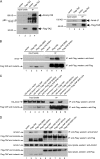
FIGURE 1.
FIGURE 1.
Interaction of TAZ and YAP with Amot and AmotL1 is dependent of the WW domain and the PPXY. A, lysates derived from HEK293 cells transfected with vector or FLAG-TAZ expression construct were immunoprecipitated with FLAG antibodies. The precipitates and lysates were analyzed by immunoblot to detect FLAG-TAZ and co-precipitated Amot (left panel) and AmotL1 (right panel). The p130 and p80 forms of Amot were indicated. B, lysates derived from HEK293 cells transfected with the indicated expression constructs were immunoprecipitated with FLAG antibodies. The precipitates and lysates were analyzed by immunoblot to detect FLAG-tagged TAZ proteins (lower panel) and co-precipitated endogenous Amot (top panel). C, lysates derived from HEK293 cells transfected with the indicated expression constructs were immunoprecipitated with FLAG antibodies. The precipitates and lysates were analyzed by immunoblot to detect FLAG-tagged YAP proteins (lower panel) and co-precipitated HA-Amot (top panel). D, lysates derived from HEK293 cells transfected with the indicated expression constructs were immunoprecipitated with FLAG antibodies. The precipitates were analyzed by immunoblot to detect FLAG-tagged proteins (second panel) and co-precipitated endogenous AmotL1 (top panel). The input lysates were analyzed for detection of AmotL1 (third panel) and FLAG-tagged protein (bottom panel). E, lysates derived from cells expressing HA-Amot and its indicated mutants were immunoprecipitated with anti-HA antibodies. The precipitates (upper panels) and lysates (lower panels) were processed for immunoblot to detect HA-Amot and co-recovered endogenous TAZ/YAP. F–H, direct interaction of PPXY motif-containing Amot fragment with the YAP-WW1+2 domain. F, the top panel is the raw heat response obtained after injection of YAP-WW1+2 domain into ITC cell containing the N-terminal fragment of Amot. The bottom panel reflects the integrated peak areas normalized to moles of YAP-WW1+2, and the solid line is the least-squares fit to the binding isotherm. The affinity is 9 ± 1 μm. G, no significant heat response and binding when YAP-WW1+2m was injected. In this mutant, the tryptophan in the binding site of both WW domains is mutated to alanine. H, the affinity between the N-terminal fragment of Amot and various WW domains are tabulated. aa, amino acids.
FIGURE 1.
FIGURE 1.
Interaction of TAZ and YAP with Amot and AmotL1 is dependent of the WW domain and the PPXY. A, lysates derived from HEK293 cells transfected with vector or FLAG-TAZ expression construct were immunoprecipitated with FLAG antibodies. The precipitates and lysates were analyzed by immunoblot to detect FLAG-TAZ and co-precipitated Amot (left panel) and AmotL1 (right panel). The p130 and p80 forms of Amot were indicated. B, lysates derived from HEK293 cells transfected with the indicated expression constructs were immunoprecipitated with FLAG antibodies. The precipitates and lysates were analyzed by immunoblot to detect FLAG-tagged TAZ proteins (lower panel) and co-precipitated endogenous Amot (top panel). C, lysates derived from HEK293 cells transfected with the indicated expression constructs were immunoprecipitated with FLAG antibodies. The precipitates and lysates were analyzed by immunoblot to detect FLAG-tagged YAP proteins (lower panel) and co-precipitated HA-Amot (top panel). D, lysates derived from HEK293 cells transfected with the indicated expression constructs were immunoprecipitated with FLAG antibodies. The precipitates were analyzed by immunoblot to detect FLAG-tagged proteins (second panel) and co-precipitated endogenous AmotL1 (top panel). The input lysates were analyzed for detection of AmotL1 (third panel) and FLAG-tagged protein (bottom panel). E, lysates derived from cells expressing HA-Amot and its indicated mutants were immunoprecipitated with anti-HA antibodies. The precipitates (upper panels) and lysates (lower panels) were processed for immunoblot to detect HA-Amot and co-recovered endogenous TAZ/YAP. F–H, direct interaction of PPXY motif-containing Amot fragment with the YAP-WW1+2 domain. F, the top panel is the raw heat response obtained after injection of YAP-WW1+2 domain into ITC cell containing the N-terminal fragment of Amot. The bottom panel reflects the integrated peak areas normalized to moles of YAP-WW1+2, and the solid line is the least-squares fit to the binding isotherm. The affinity is 9 ± 1 μm. G, no significant heat response and binding when YAP-WW1+2m was injected. In this mutant, the tryptophan in the binding site of both WW domains is mutated to alanine. H, the affinity between the N-terminal fragment of Amot and various WW domains are tabulated. aa, amino acids.
FIGURE 2.
FIGURE 2.
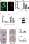
Amot and AmotL1 inhibit TAZ transcriptional outcome and oncogenic property in promoting anchorage-independent growth. A, MCF7 cells were co-transfected with FLAG-TAZ-S89A and HA-Amot. Cells were then processed to detect the expressed proteins using mouse anti-FLAG and rabbit anti-HA antibodies followed by secondary antibodies (green and red for FLAG and HA tag, respectively). FLAG-TAZ-S89A, expressed alone, is essentially in the nucleus (cell 1). When Amot was expressed at low moderate levels, some S89A was shifted to the cytoplasm (cell 2), and S89A was largely detected in the cytoplasm when Amot was expressed at high levels (cells 3 and 4). B, the mRNA levels of endogenous CTGF gene were measured by real-time PCR in cells transfected with vector (columns 1–4) or TAZ-89A-expressing construct (columns 5–8) together with the expression vectors indicated at the bottom (ctrl, vector control; A, Amot coding cDNA; L1, AmotL1 coding cDNA; A+L1, Amot and AmotL1 coding cDNAs together). The levels of CTGF mRNA were normalized to that detected in column 1, which was arbitrarily set at 1. C, the mRNA levels of Cyr61 were measured in those cells described in panel B. The Cyr61 mRNA levels were normalized to that detected in column 1, which was arbitrarily set at 1. D, the expression of S89A, Amot, or AmotL1 was examined by immunoblot. E, NIH3T3 cells were transduced to express TAZ-S89A (left panel) or YAP-S127A (right panel) along with vector (upper panels) or with Amot-expressing construct (bottom panels). Cells were grown in soft agar, and colonies were stained and photographed. F, the quantitative results derived from three independent experiments similar to D were shown. Error bars in panels B, C, and F indicate S.E.
FIGURE 3.
FIGURE 3.
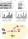
Endogenous Amot but not AmotL1 negatively regulates the expression of endogenous CTGF and Cyr61 genes. A, the lysates derived from the indicated nine different human cell lines were analyzed by immunoblot to detect endogenous Amot (upper panel) and AmotL1 (lower panel). β-Actin was used as control. B, Amot and AmotL1 protein levels in cells transfected with their respective siRNA were determined by immunoblot. C, the mRNA levels of CTGF were measured by real-time PCR in HEK293 cells transfected with vector (columns 1–4) or TAZ-89A-expressing construct (columns 5–8) together with siRNA indicated at the bottom (ctrl siRNA, control siRNA; A siRNA, Amot siRNA; L1 siRNA, AmotL1 siRNA; A+L1 siRNA, Amot and AmotL1 siRNA together). The levels of CTGF mRNA were normalized to that detected in column 1, which was arbitrarily set at 1. D, the mRNA levels of Cyr61 were measured in those cells described in panel C. The Cyr61 mRNA levels were normalized to that detected in column 1, which was arbitrarily set at 1. Error bars in panels C and D indicate S.E. E, a working model for diverse regulatory mechanisms for TAZ and YAP. The Hippo pathway causes cytoplasmic sequestration of TAZ and YAP through phosphorylation of Ser89 and Ser127, respectively. Furthermore, Hippo pathway-mediated phosphorylation of Ser314 and Ser381 leads to further phosphorylation, ubiquitination, and proteasomal degradation of TAZ and YAP, respectively. Interaction with TEADs is important for nuclear accumulation and transcriptional outcome of TAZ and YAP. The results presented in this study suggest that Amot and AmotL1 (likely also AmotL2) function as negative regulators of TAZ and YAP through direct interaction with the WW domain of TAZ and the first WW domain of YAP via the first PPXY motif of Amot and AmotL1 that is also conserved in AmotL2, leading to their cytoplasmic retention.

References

    1. Chan S. W., Lim C. J., Chen L., Chong Y. F., Huang C., Song H., Hong W. (October 13, 2010) J. Cell. Physiol. 10.1002/JCP.22435 - DOI - PubMed
    1. Zeng Q., Hong W. (2008) Cancer Cell 13, 188–192 - PubMed
    1. Pan D. (2010) Dev. Cell 19, 491–505 - PMC - PubMed
    1. Zhao B., Li L., Lei Q., Guan K. L. (2010) Genes Dev. 24, 862–874 - PMC - PubMed
    1. Grusche F. A., Richardson H. E., Harvey K. F. (2010) Curr. Biol. 20, R574–R582 - PubMed

Publication types

MeSH terms