. 2011 Mar 1;50(8):1359-67.
doi: 10.1021/bi101749s. Epub 2011 Jan 31.

The Cullin-RING E3 ubiquitin ligase CRL4-DCAF1 complex dimerizes via a short helical region in DCAF1

Affiliations

The Cullin-RING E3 ubiquitin ligase CRL4-DCAF1 complex dimerizes via a short helical region in DCAF1

Jinwoo Ahn et al. Biochemistry. .

Abstract

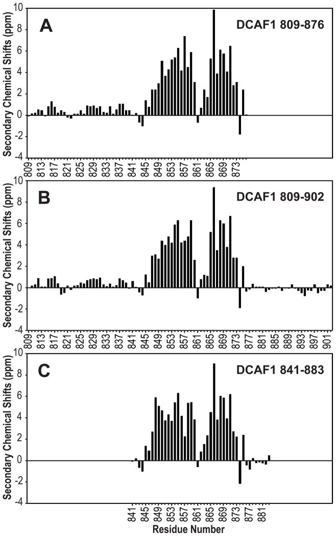
The cullin4A-RING E3 ubiquitin ligase (CRL4) is a multisubunit protein complex, comprising cullin4A (CUL4), RING H2 finger protein (RBX1), and DNA damage-binding protein 1 (DDB1). Proteins that recruit specific targets to CRL4 for ubiquitination (ubiquitylation) bind the DDB1 adaptor protein via WD40 domains. Such CRL4 substrate recognition modules are DDB1- and CUL4-associated factors (DCAFs). Here we show that, for DCAF1, oligomerization of the protein and the CRL4 complex occurs via a short helical region (residues 845-873) N-terminal to DACF1's own WD40 domain. This sequence was previously designated as a LIS1 homology (LisH) motif. The oligomerization helix contains a stretch of four Leu residues, which appear to be essential for α-helical structure and oligomerization. In vitro reconstituted CRL4-DCAF1 complexes (CRL4(DCAF1)) form symmetric dimers as visualized by electron microscopy (EM), and dimeric CRL4(DCAF1) is a better E3 ligase for in vitro ubiquitination of the UNG2 substrate compared to a monomeric complex.


Figures

Figure 1
Figure 1. DCAF1 oligomerizes via a putative LisH motif IN VIVO
A. N-terminally FLAG-tagged and C-terminally V5-tagged DCAF1 (FLAG-DCAF1-V5, residues 96–1507) was transiently co-expressed with N-terminally Myc-tagged and C-terminally V5-tagged DCAF1 (Myc-DCAF1-V5, residues 96–1507) in SF9 insect cells. Cell lysates were immunoprecipitated with anti-FLAG agarose affinity gel, and pull-down mixtures were analyzed by immunoblotting with anti-FLAG and anti-Myc antibodies (IP), after separation by SDS-PAGE and transfer to nitrocellulose. 5% of total cell lysates were also immunoblotted with anti-V5 antibody (Input). B. FLAG-DCAF1-V5 was transiently co-expressed in SF9 cells with several deletion constructs of DCAF1 (residues 572–1507, 817–1507, 876–1396, 987–1396, and 1005–1507), containing the V5-tag at their C-termini. Each cell lysate was subjected to immunoprecipitation with anti-FLAG agarose affinity gel, and pull-down mixtures were analyzed by immunoblotting with anti-V5 antibody. Constructs pulled-down by FLAG-DCAF1-V5 are labeled by (+), and those not pulled down by (-).
Figure 2
Figure 2. Secondary structure characterization of isolated DCAF1 peptides by NMR
A–C, Secondary chemical shifts, ΔCα minus ΔCβ, (48) for DCAF1 809–876 (A), 809–902 (B) and 841–883 (C) are shown along the amino acid sequence. ΔCα and ΔCβ values were calculated for all assigned resonances by subtracting measured Cα and Cβ shifts from random coil values (47).
Figure 3
Figure 3. The four Leu region is critical for oligomerization of DCAF1
A. Sequence alignment of putative LisH domains in orthologous DCAF1s. Identical amino acid residues are underlined and the LisH motif, L-X2-L-X3–5-L-X3–5-L is displayed at the bottom. X represents any amino acid. B. FLAG-DCAF1-V5 (encoding residues of 96–1507) was transiently co-expressed with the N-terminally deleted DCAF1 (ΔN-DCAF1-V5, residues of 817–1507), site-directed mutants ΔN-DCAF1 (1LL/EE)-V5, and ΔN-DCAF1 (2LL/EE)-V5, respectively. 1LL/EE corresponds to the Leu850Glu, Leu851Glu double mutant, and 2LL/EE to the Leu852Glu, Leu853Glu double mutant. Cell lysates were immunoprecipitated with anti-FLAG agarose affinity gel. Proteins bound to antibodies were separated by SDS-PAGE, and subjected to immunoblotting with anti-V5 antibody after transfer to nitrocellulose. C. The DCAF1 region corresponding to residues 809–876 and the 1LL/EE and 2LL/EE mutants thereof were expressed and purified from E. coli as thioredoxin (Trx) fusion proteins. Trx and the three Trx-DCAF1 proteins were separated by SDS-PAGE and stained with Coomassie Brillant Blue. D. Multi-angle light scattering of purified Trx-DCAF1 fusion proteins and Trx as a control. Each protein (ca. 2 mg/mL) was injected into an analytical Superdex200 gel filtration column at a flow rate of 0.5 mL/min. The UV (A280) elution profiles of Trx-DCAF1 809–876 WT (▼), Trx-DCAF1 809–876 1LL/EE (●), Trx-DCAF1 809–876 2LL/EE (◆), and Trx (■) are shown. The estimated molecular masses from the scattering data are shown across the elution peaks.
Figure 4
Figure 4. Secondary structure characterization of WT and mutant LisH motif peptides by CD
Circular Dichroism (CD) spectra of WT DCAF1 809–876 (1, ●), 1LL/EE 809–876 (2, ▲), and 2LL/EE 809–876 (3, ■), recorded at 3.3 μM in a buffer containing 2.5 mM sodium phosphate, pH 7.5, 15 mM NaCl, at 25 °C. Data were collected at 0.5 nm intervals, scanned from 250–195 nm, and averaged over 10 measurements. The inset shows the SDS-PAGE analysis of the peptides used for CD.
Figure 5
Figure 5. Characterization of the DDB1-DCAF1 complex by SEC-MALS and SDS-PAGE
The DDB1-DCAF1 817–1507 protein complex was injected into an analytical gel filtration column at a flow rate of 0.5 mL/min. The UV (A280) elution profile and the estimated molecular mass from the scattering data are shown across the elution peaks. The protein complex was separated by SDS-PAGE and proteins visualized by Coomassie Brillant Blue.
Figure 6
Figure 6. Electron microscopy of CRL4DCAF1 E3 ligase complexes
A. SDS-PAGE analysis of the CRL4DCAF1 E3 ligase complex, assembled with CUL4A, RBX1, DDB1, and DCAF1 (residues 817–1507). Each component of the complex is indicated. B. SEC-MALS analysis of the CRL4DCAF1 E3 ligase complex. The protein complex was injected into an analytical gel filtration column at a flow rate of 0.5 mL/min and the data were analyzed as described in Experimental Procedures. The estimated molecular mass is shown across the elution peak. C. Electron micrograph of negatively stained CRL4DCAF1 complexes (scale bar, 100 nm). D. Selected 2D class averages of CRL4DCAF1 complex particles selected from the micrograph. Some class averages show near mirror symmetry suggesting the presence of a 2-fold axis in the complexes.
Figure 7
Figure 7. Ubiquitination activity of monomeric and dimeric CRL4DCAF1
A. 20 pmol of monomeric CRL4 complex bound to DCAF1 (1005–1507) and dimeric CRL4 bound to DCAF1 (817–1507) separated by SDS-PAGE and visualized with Coomassie Blue. B. Time course of UNG2 ubiquitination with dimeric CRL4DCAF1 and monomeric CRL4DCAF1 at two different concentrations (0.2 and 0.4 μM). The reaction buffer contained E1 (UBA1), E2 (UbcH5b), FLAG-tagged ubiquitin (Ub), and NusA-Vpr-ΔC. Ubiquitinated proteins were detected by immunoblotting with either anti-UNG2 or anti-FLAG antibody after separation of the reaction mixture on SDS-PAGE and transfer to nitrocellulose.

References

    1. Goldstein G, Scheid M, Hammerling U, Schlesinger DH, Niall HD, Boyse EA. Isolation of a polypeptide that has lymphocyte-differentiating properties and is probably represented universally in living cells. Proc Natl Acad Sci USA. 1975;72:11–15. - PMC - PubMed
    1. Ciechanover A, Finley D, Varshavsky A. Ubiquitin dependence of selective protein degradation demonstrated in the mammalian cell cycle mutant ts85. Cell. 1984;37:57–66. - PubMed
    1. Ciechanover A, Finley D, Varshavsky A. The ubiquitin-mediated proteolytic pathway and mechanisms of energy-dependent intracellular protein degradation. J Cell Biochem. 1984;24:27–53. - PubMed
    1. Hershko A, Ciechanover A. The ubiquitin system. Annu Rev Biochem. 1998;67:425–479. - PubMed
    1. Pickart CM. Mechanisms underlying ubiquitination. Annu Rev Biochem. 2001;70:503–533. - PubMed

Publication types

MeSH terms