

Review
doi: 10.1146/annurev-med-041709-133809.
Imaging of atherosclerosis
Affiliations
- PMID: 21226610
- PMCID: PMC4041162
- DOI: 10.1146/annurev-med-041709-133809
Item in Clipboard
Review
Imaging of atherosclerosis
Annu Rev Med.
2011.
Abstract
It is now well recognized that the atherosclerotic plaques responsible for thrombus formation are not necessarily those that impinge most on the lumen of the vessel. Nevertheless, clinical investigations for atherosclerosis still focus on quantifying the degree of stenosis caused by plaques. Many of the features associated with a high-risk plaque, including a thin fibrous cap, large necrotic core, macrophage infiltration, neovascularization, and intraplaque hemorrhage, can now be probed by novel imaging techniques. Each technique has its own strengths and drawbacks. In this article, we review the various imaging modalities used for the evaluation and quantification of atherosclerosis.
Figures
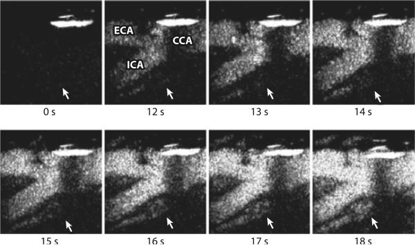
Contrast-enhanced ultrasound of the carotid plaque. Time 0 s shows the total subtracted image (black) in the nonlinear imaging mode at the carotid bifurcation before the arrival of the contrast agent. From time 12 s, the enhancement of the carotid lumen (white) is evident. Note the subsequent enhancement of the plaque neovascularization from time 14 s (white arrow). CCA, common carotid artery; ECA, external carotid artery; ICA, internal carotid artery. Reproduced with permission from Prof. Ed Leen, Imperial College London.
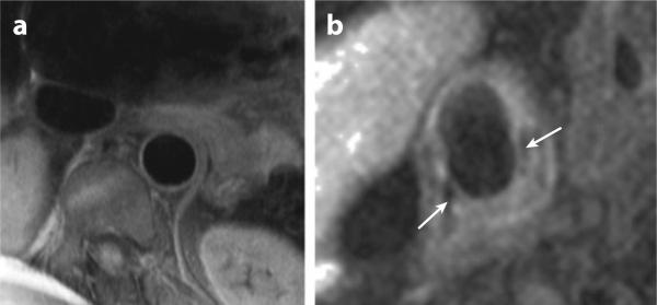
Magnetic resonance imaging of the aorta and carotid artery. (a) The descending thoracic aorta adjacent to the lumbar spine. The vessel wall is clearly demarcated and can be seen to be normal. (b) Complicated right carotid plaque with a necrotic lipid core and a thin fibrous cap (white arrows). Images courtesy of Dr. Robin Choudhury.
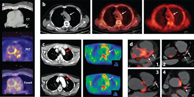
(a) [18F]FDG PET/CT image (CT/PET/Fused) of aorta. Image courtesy of Prof. Fayad. (b) [11C]-choline PET/CT images of the aortic arch: CT (left), PET (middle), and fused (right). Tracer uptake coincides with calcification (arrows indicate calcification site). (c) [11C]PK11195 CT angiogram (left), PET image (right). Patient with active giant cell arteritis with increased tracer uptake in the lateral aspect of the aortic arch (arrow). The CT angiography demonstrates corresponding thickening of the aortic wall (arrow). (d ) Coregistered [18F]FDG PET/CT images show FDG uptake within specified coronary vascular locations (85). Arrows show stent locations. Hatched arrows show lesions within the left main coronary artery (LM). (d1) Increased uptake in both LM and stented culprit lesions in a subject presenting with acute coronary syndrome (ACS). (d2) More modest uptake in a coronary lesion that was recently stented for a stable coronary syndrome (some FDG uptake is also noted in a mixed plaque within the LM). (d3) Modest FDG uptake within a lesion stented several months before imaging. (d4) The FDG uptake at the trifurcation of the LM in a subject presenting with ACS.
References
-
- Lloyd-Jones D, Adams RJ, Brown TM, et al. Heart disease and stroke statistics—2010 update: a report from the American Heart Association. Circulation. 2010;121(7):e46–e215. - PubMed
-
- Hellings WE, Peeters W, Moll FL, et al. From vulnerable plaque to vulnerable patient: the search for biomarkers of plaque destabilization. Trends Cardiovasc. Med. 2007;17(5):162–71. - PubMed
-
- Hackett D, Davies G, Maseri A. Pre-existing coronary stenoses in patients with first myocardial infarction are not necessarily severe. Eur. Heart J. 1988;9(12):1317–23. - PubMed
-
- Randomised trial of cholesterol lowering in 4444 patients with coronary heart disease: the Scandinavian Simvastatin Survival Study (4S). Lancet. 1994;344(8934):1383–89. - PubMed
-
- Topol EJ, Nissen SE. Our preoccupation with coronary luminology. The dissociation between clinical and angiographic findings in ischemic heart disease. Circulation. 1995;92(8):2333–42. - PubMed
Publication types
MeSH terms
Substances
Grants and funding
LinkOut - more resources
Full Text Sources
Other Literature Sources
Medical
Miscellaneous
