

Mesenchymal stem cell derived hematopoietic cells are permissive to HIV-1 infection
- PMID: 21226936
- PMCID: PMC3025950
- DOI: 10.1186/1742-4690-8-3
Mesenchymal stem cell derived hematopoietic cells are permissive to HIV-1 infection
Abstract
Background: Tissue resident mesenchymal stem cells (MSCs) are multipotent, self-renewing cells known for their differentiation potential into cells of mesenchymal lineage. The ability of single cell clones isolated from adipose tissue resident MSCs (ASCs) to differentiate into cells of hematopoietic lineage has been previously demonstrated. In the present study, we investigated if the hematopoietic differentiated (HD) cells derived from ASCs could productively be infected with HIV-1.
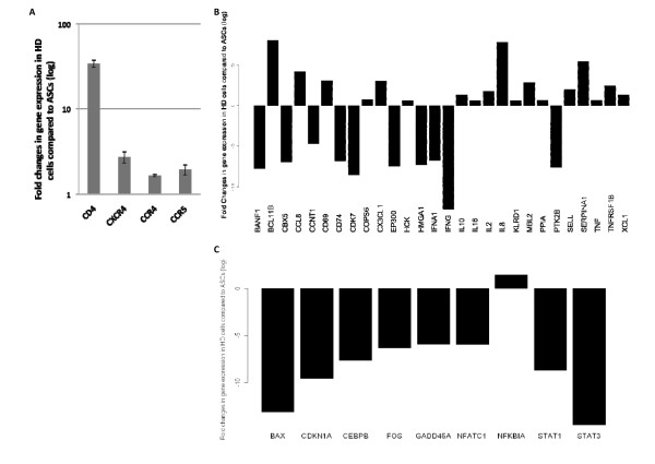
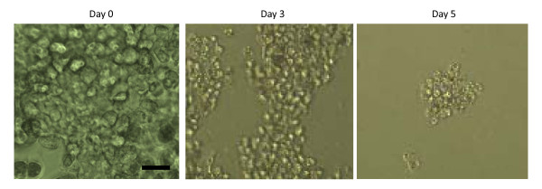
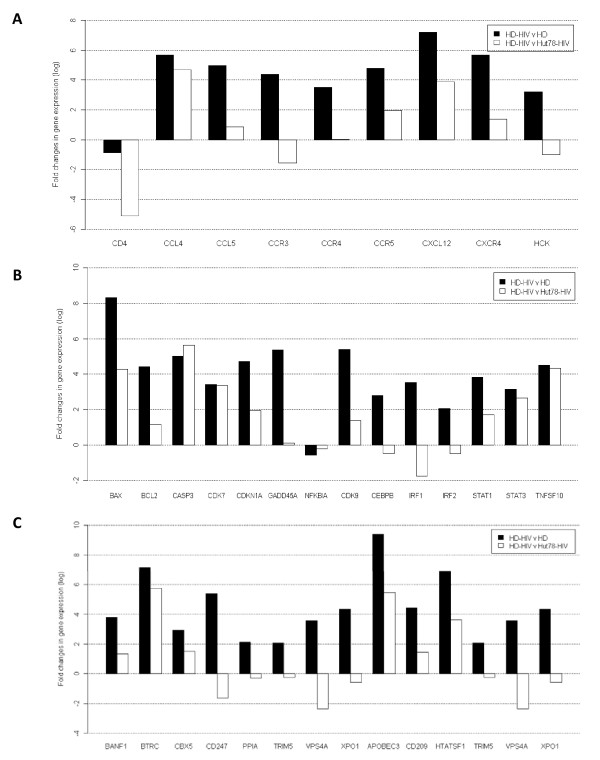
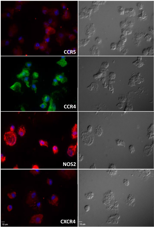
Results: HD cells were generated by differentiating clonally expanded cultures of adherent subsets of ASCs (CD90+, CD105+, CD45-, and CD34-). Transcriptome analysis revealed that HD cells acquire a number of elements that increase their susceptibility for HIV-1 infection, including HIV-1 receptor/co-receptor and other key cellular cofactors. HIV-1 infected HD cells (HD-HIV) showed elevated p24 protein and gag and tat gene expression, implying a high and productive infection. HD-HIV cells showed decreased CD4, but significant increase in the expression of CCR5, CXCR4, Nef-associated factor HCK, and Vpu-associated factor BTRC. HIV-1 restricting factors like APOBEC3F and TRIM5 also showed up regulation. HIV-1 infection increased apoptosis and cell cycle regulatory genes in HD cells. Although undifferentiated ASCs failed to show productive infection, HIV-1 exposure increased the expression of several hematopoietic lineage associated genes such as c-Kit, MMD2, and IL-10.
Conclusions: Considering the presence of profuse amounts of ASCs in different tissues, these findings suggest the possible role that could be played by HD cells derived from ASCs in HIV-1 infection. The undifferentiated ASCs were non-permissive to HIV-1 infection; however, HIV-1 exposure increased the expression of some hematopoietic lineage related genes. The findings relate the importance of ASCs in HIV-1 research and facilitate the understanding of the disease process and management strategies.
Figures


References
-
- Perelson AS, Essunger P, Ho DD. Dynamics of HIV-1 and CD4+ lymphocytes in vivo. AIDS. 1997;11(Suppl A):S17–24. - PubMed
-
- Ogg GS, Jin X, Bonhoeffer S, Moss P, Nowak MA, Monard S, Segal JP, Cao Y, Rowland-Jones SL, Hurley A, Markowitz M, Ho DD, McMichael AJ, Nixon DF. Decay kinetics of human immunodeficiency virus-specific effector cytotoxic T lymphocytes after combination antiretroviral therapy. J Virol. 1999;73:797–800. - PMC - PubMed
-
- Moore JP, Kitchen SG, Pugach P, Zack JA. The CCR5 and CXCR4 coreceptors--central to understanding the transmission and pathogenesis of human immunodeficiency virus type 1 infection. AIDS Res Hum Retroviruses. 2004;20:111–126. - PubMed
-
- Zaitseva M, Peden K, Golding H. HIV coreceptors: role of structure, posttranslational modifications, and internalization in viral-cell fusion and as targets for entry inhibitors. Biochim Biophys Acta. 2003;1614:51–61. - PubMed
-
- Ruiz ME, Cicala C, Arthos J, Kinter A, Catanzaro AT, Adelsberger J, Holmes KL, Cohen OJ, Fauci AS. Peripheral blood-derived CD34+ progenitor cells: CXC chemokine receptor 4 and CC chemokine receptor 5 expression and infection by HIV. J Immunol. 1998;161:4169–4176. - PubMed
Publication types
MeSH terms
Substances
Grants and funding
LinkOut - more resources
Full Text Sources
Other Literature Sources
Medical
Research Materials
Miscellaneous
