. 2011 Jan 12;31(2):402-13.
doi: 10.1523/JNEUROSCI.2897-10.2011.

FoxG1 promotes the survival of postmitotic neurons

Affiliations

FoxG1 promotes the survival of postmitotic neurons

Somasish Ghosh Dastidar et al. J Neurosci. .

Abstract

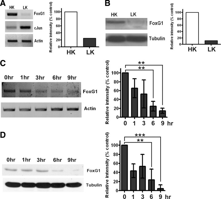
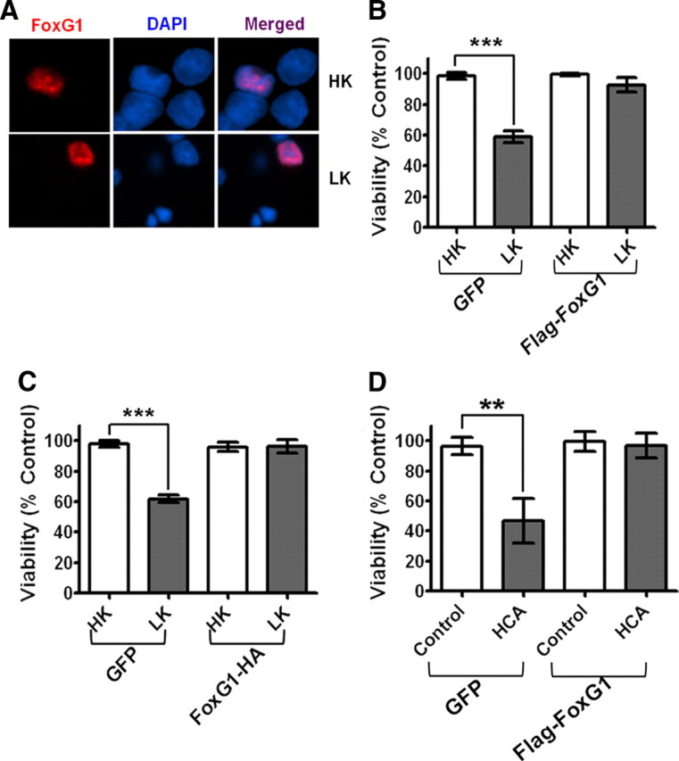
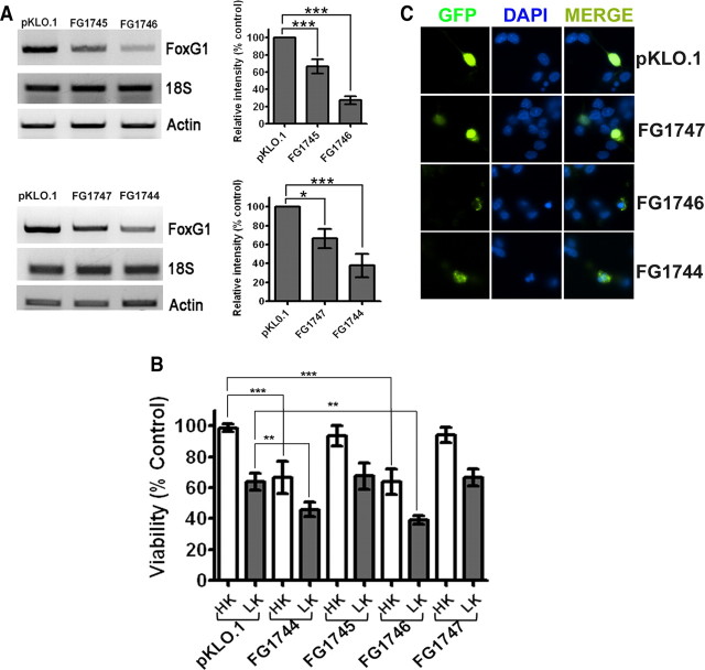
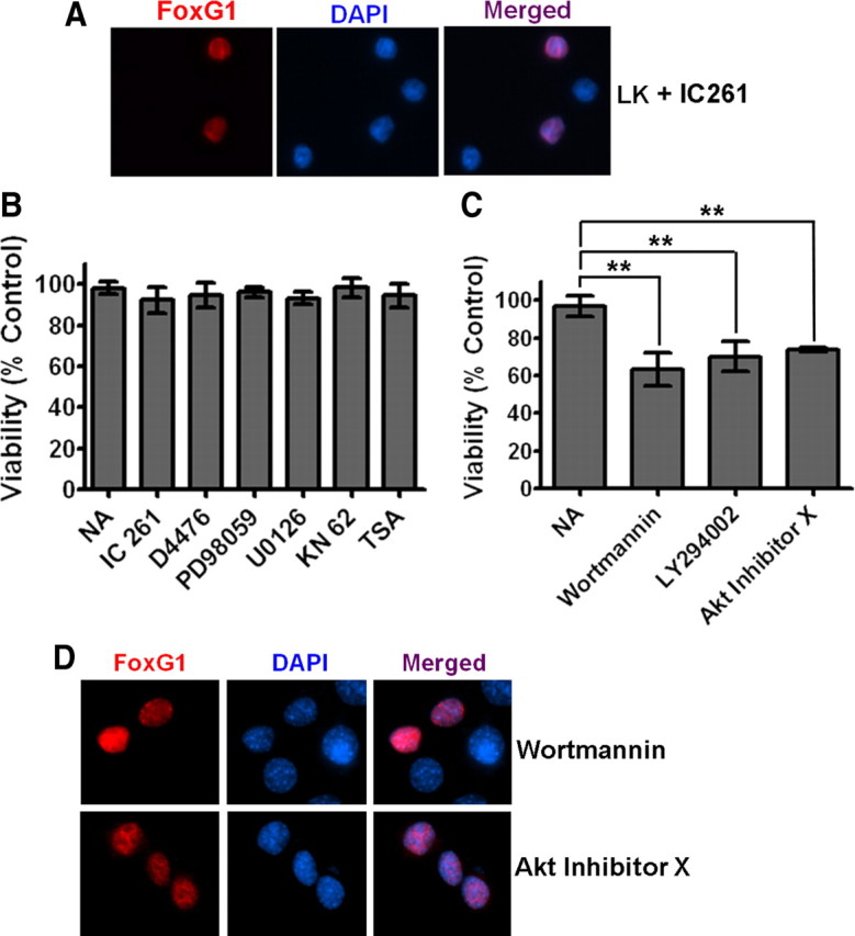
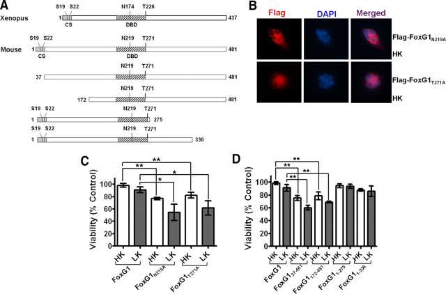
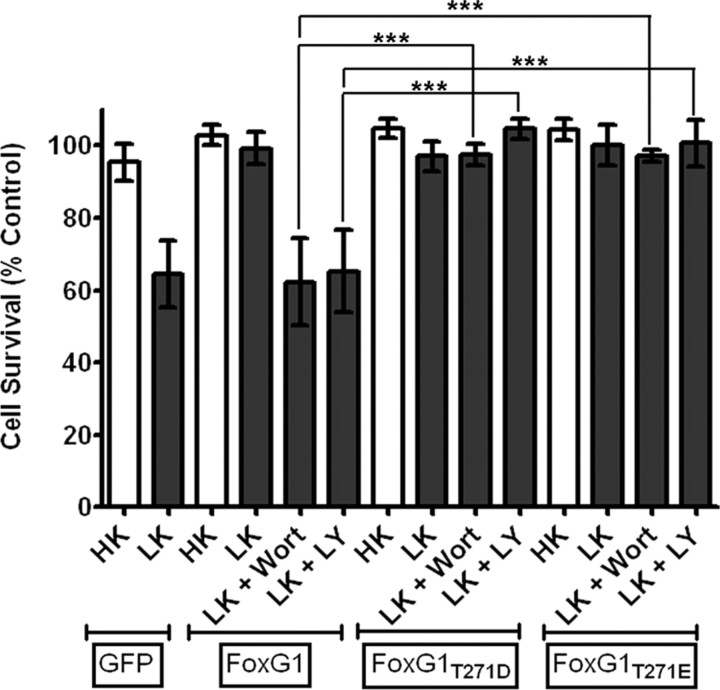
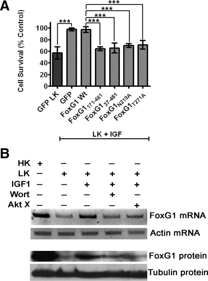
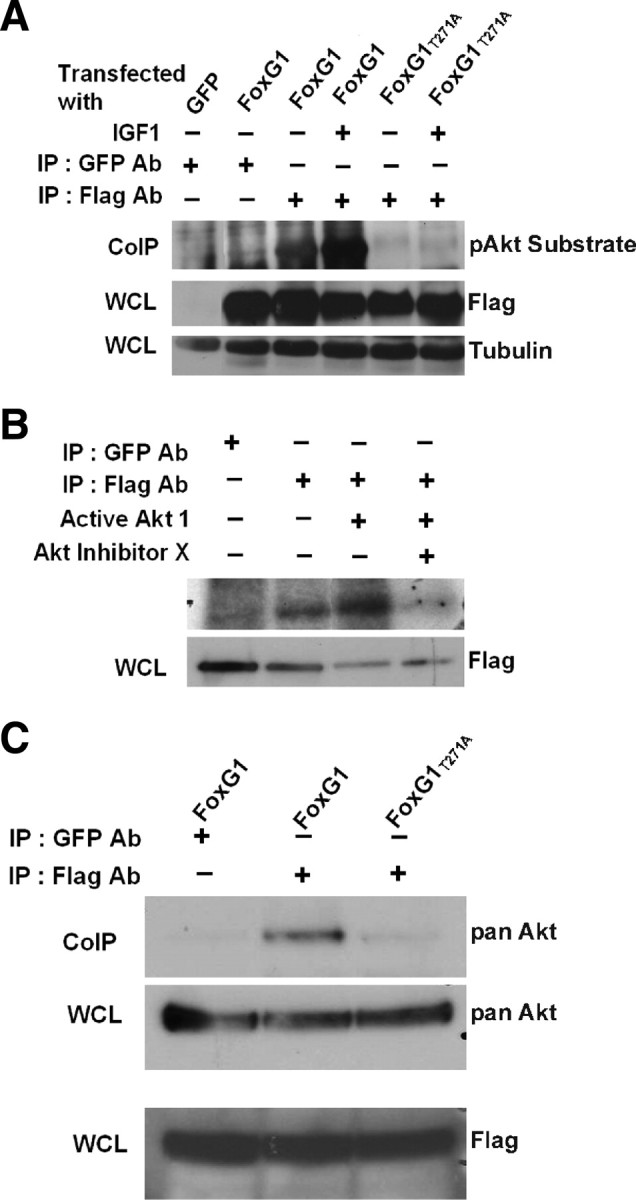
The transcription factor FoxG1 regulates neurogenesis in the embryonic telencephalon as well as a number of other neurodevelopmental processes. While FoxG1 continues to be expressed in neurons postnatally and through adulthood, its role in fully differentiated neurons is not known. The current study demonstrates that FoxG1 promotes the survival of postmitotic neurons. In cerebellar granule neurons primed to undergo apoptosis, FoxG1 expression is reduced. Ectopic expression of FoxG1 blocks neuronal death, whereas suppression of its expression induces death in otherwise healthy neurons. The first 36 residues of FoxG1 are necessary for its survival-promoting effect, while the C-terminal half of the protein is dispensable. Mutation of Asp219, a residue necessary for DNA binding, abrogates survival promotion by FoxG1. Survival promotion is also eliminated by mutation of Thr271, a residue phosphorylated by Akt. Pharmacological inhibition of Akt blocks the survival effects of wild-type FoxG1 but not forms in which Thr271 is replaced with phosphomimetic residues. Treatment of neurons with IGF-1, a neurotrophic factor that promotes neuronal survival by activating Akt, prevents the apoptosis-associated downregulation of FoxG1 expression. Moreover, the overexpression of dominant-negative forms of FoxG1 blocks the ability of IGF-1 to maintain neuronal survival suggesting that FoxG1 is a downstream mediator of IGF-1/Akt signaling. Our study identifies a new and important function for FoxG1 in differentiated neurons.


Figures

Figure 1.
Figure 1.
Expression of FoxG1 in postmitotic neurons. A, RNA isolated from CGNs treated with HK or LK for 6 h were subjected to RT-PCR analysis of FoxG1 expression. FoxG1 mRNA expression is reduced at 6 h of LK treatment. In contrast, the expression of the proapoptotic c-Jun gene is induced in neurons primed to die by LK treatment. Actin serves as the loading control. B, Whole-cell lysates were prepared from CGNs treated with HK or LK for 6 h and subjected to Western blot analysis using a FoxG1 antibody. FoxG1 is downregulated in LK. Tubulin serves as the loading control. C, RNA isolated from CGNs treated with LK for 0, 1, 3, 6, and 9 h was subjected to RT-PCR analysis of FoxG1 expression. Actin served as loading control. Densitometric analysis indicates reduction of FoxG1 mRNA level with time. Results were obtained from three separate experiments. **p < 0.01. For statistical analysis, one-way ANOVA was performed using Bonferroni's multiple-comparison test. D, Whole-cell lysates prepared from CGNs treated with LK for 0, 1, 3, 6, and 9 h were subjected to Western blot analysis using a FoxG1 antibody. Tubulin serves as the loading control. Densitometric analysis indicated reduction of FoxG1 protein level with time. Results were obtained from three separate experiments. ***p < 0.001, **p < 0.01. For statistical analysis, one-way ANOVA was performed using Bonferroni's multiple-comparison test.
Figure 2.
Figure 2.
Effect of FoxG1 overexpression in postmitotic neuronal survival: CGN cultures were transfected with GFP and Flag-FoxG1 or FoxG1-HA for 12 h and then switched to HK or LK medium for 24 h followed by immunocytochemistry. The viability of transfected neurons was quantified by DAPI staining and normalized to GFP-transfected cells in HK. FoxG1 protects neurons independent of its tag. A, Localization of ectopically expressed FoxG1 in CGNs. Transfected neurons were identified by immunocytochemistry using Flag antibody. FoxG1 localizes exclusively in the nucleus in HK- and LK-treated neurons. B, FoxG1 protects CGNs from LK-induced apoptosis. C, CGN cultures transfected with either GFP or FoxG1-HA. FoxG1-HA protects neurons from LK-induced apoptosis. Results were obtained from three separate experiments done in duplicate. ***p < 0.001 as compared to control (GFP-transfected neurons in HK). D, Two-day-old cultures of primary cortical neurons transfected with GFP or Flag FoxG1 were treated with either medium having no additives (NA) or medium containing 1 mm HCA for 20 h. Viability of transfected neurons was quantified after immunocytochemistry using GFP or Flag antibody and normalized to the viability of control cultures (GFP-transfected cells receiving no additives). **p < 0.01 as compared to GFP-transfected cortical neurons with NA.
Figure 3.
Figure 3.
Suppression of FoxG1 expression promotes neuronal death. CGN cultures were cotransfected with plasmids expressing GFP and either pKLO.1 or one of the four different FoxG1 shRNAs (FG1744, FG1745, FG1746, and FG1747). The ability of the shRNAs to suppress FoxG1 expression was evaluated in HT22 cells, and their effect on neuronal survival was analyzed in CGNs. A, RNA was extracted from HT22 cells 72 h after transfection with shRNA constructs and subjected to RT-PCR analysis using primers against FoxG1, 18S ribosomal RNA, and actin. Results for the four FoxG1 shRNAs [FG1745 and FG1746 (upper panel) and FG1747 and FG1744 (lower panel)] along with the control (pKLO.1) are shown. Densitometric analysis of the FoxG1 band from three separate experiments was performed using Kodak 1D software, and the results are shown in the graph. ***p < 0.001, *p < 0.05. For statistical analysis, one-way ANOVA was performed using Bonferroni's multiple-comparison test. B, CGNs were cotransfected with plasmids expressing GFP and pKLO.1 or one of the four FoxG1 shRNAs and treated with HK or LK medium. Results come from three separate experiments performed in duplicate. ***p < 0.001 as compared to neurons cotransfected with GFP and pKLO.1 in HK. **p < 0.01 as compared to neurons cotransfected with GFP and pKLO.1 in LK. C, Appearance of neurons transfected with pKLO.1 plasmid or FoxG1 shRNA (FG1747, FG1746, FG1744). Transfected neurons were identified by GFP immunocytochemistry and nuclei stained with DAPI.
Figure 4.
Figure 4.
The PI-3 kinase/Akt pathway mediates the anti-apoptotic effect of FoxG1. A, Ectopically expressed FoxG1 was detected immunocytochemically with a Flag antibody and nuclei were stained with DAPI. The figure shows nuclear localization of FoxG1 in neurons treated with LK in presence of IC261. CKI inhibition does not alter the nuclear localization of FoxG1. B, CGN cultures transfected with Flag-FoxG1 were switched to LK medium having no additives (NA), LK medium containing 10 μm IC261, 10 μm D4476, 40 μm PD98059, 10 μm U0126, 50 μm KN62, or 1 μm trichostatin A. The survival status of transfected neurons was quantified 24 h later by Flag immunocytochemistry and DAPI nuclear staining. Inhibitors against CKI, CaMK, HDACs, and the RAf-MEK-ERK pathway did not have any effect on FoxG1-mediated survival. Results were obtained from three independent experiments performed in duplicate. Control experiments (supplemental Table 1, available at www.jneurosci.org as supplemental material) confirmed that all the pharmacological agents used in this analysis inhibit the activities of their targets. C, CGN cultures transfected with Flag-FoxG1 were switched to LK medium with NA or LK medium containing 200 nm wortmannin, 20 μm LY294002, and 5 μm Akt inhibitor X. Inhibitors of the PI-3 kinase/Akt pathway blocked FoxG1 from helping CGNs to survive in LK conditions. **p < 0.01 as compared to FoxG1-transfected CGNs treated with LK. Results were obtained from three independent experiments performed in duplicates. D, HT22 cells were transfected with Flag-FoxG1. Six hours after transfection, the medium was changed to fresh medium supplemented with 200 nm wortmannin or 5 μm Akt inhibitor X for 12 h. Immunocytochemistry was performed with Flag antibody and nuclei were stained with DAPI. Blocking PI3K/Akt does not block nuclear translocation of FoxG1.
Figure 5.
Figure 5.
Sites or domains responsible for FoxG1 mediated neuronal survival. A, Schematic diagram of the wild-type FoxG1 protein from Xenopus [which was used by Regad et al. (2007)] and the mouse FoxG1 protein used in this study. Position of relevant amino acids as well as the conserved sequence region (CS) and the DBD is indicated. Also shown are the various FoxG1 deletion constructs used in this study. B, Immunocytochemistry result of CGN in HK transfected with Flag-FoxG1N219A and Flag-FoxG1T271A. C, CGNs were transfected with plasmids expressing wild-type FoxG1-Flag, the Akt phosphorylation site mutant Flag-FoxG1T271A, and Flag-FoxG1N219A, a mutant construct in which an aspartate residue essential for DNA binding is substituted with alanine. The neurons were then switched to HK or LK medium for 24 h, after which viability of transfected neurons was quantified. Both Flag-FoxG1N219A and Flag-FoxG1T271A failed to protect neurons from LK-induced death. Results are from three different experiments performed in duplicate. **p < 0.01 as compared to FoxG1-transfected CGNs in HK. *p < 0.05 as compared to FoxG1-transfected neurons in LK. D, Quantification of neuronal survival with the various FoxG1-Flag deletion constructs. Deletion of N-terminus regions destroys the ability of FoxG1 to prevent neuronal death in LK. Moreover, these constructs inhibit survival in HK, possibly through a dominant-negative mechanism. In contrast, C-terminus deletion constructs had no effect on HK-mediated survival. Results come from three different experiments performed in duplicate. **p < 0.01 as compared to FoxG1-transfected CGNs in HK or LK as indicated in the graph by bars.
Figure 6.
Figure 6.
Phosphorylation at Thr271 by Akt is necessary for survival-promoting activity of FoxG1. CGNs were transfected with plasmids expressing two separate phosphomimetic forms of FoxG1, Flag-FoxG1T271D, and Flag-FoxG1T271E. The cultures were then switched to HK or LK media or LK media supplemented with 200 nm wortmannin (Wort) or 20 μm LY294002 (LY). Viability of transfected neurons was quantified after immunocytochemistry and normalized to the viability of GFP-transfected neurons in HK. Survival-promoting effect of Flag-FoxG1T271D and Flag-FoxG1T271E in LK medium cannot be inhibited by inhibition of the PI-3 kinase–Akt pathway. Columns compared are indicated with bars. ***p < 0.001.
Figure 7.
Figure 7.
FoxG1 is a downstream effector of IGF-1 signaling. A, CGNs were transfected with plasmids as indicated in the graph and then switched to LK media supplemented with 25 ng/ml IGF-1. Viability of transfected neurons was quantified after immunocytochemistry using GFP/Flag antibody and normalized to the viability of GFP-transfected neurons in LK with IGF-1. IGF-1 was able to rescue GFP-transfected neurons in LK. IGF-1 was not able to rescue CGNs transfected with FoxG1 172–481 Flag, FoxG137–481-Flag, Flag-FoxG1N219A, and Flag-FoxG1T271A. ***p < 0.001 as compared to FoxG1-transfected CGNs in LK media supplemented with IGF1. Results were obtained from four independent experiments performed in duplicate. B, Six-day-old CGN cultures were treated with HK, LK, LK with IGF-1 (25 ng/ml), and LK with IGF-1 plus PI-3 kinase/Akt pathway inhibitors (wortmannin, 200 nm and Akt inhibitor X, 5 μm). RNA or protein lysates were prepared 6 h after treatment and analyzed for FoxG1 expression. Upper panel shows results of RT-PCR analysis of FoxG1 and actin mRNA. Lower panel shows results of Western blot analysis performed using FoxG1 and tubulin antibodies. IGF-1 inhibits the LK-induced reduction of FoxG1 expression. However, inhibitors of PI-3 kinase–Akt signaling inhibit the ability of IGF1 to maintain elevated FoxG1 expression.
Figure 8.
Figure 8.
FoxG1 is a substrate of Akt. A, HEK293T cells were transfected with Flag-FoxG1 or Flag-FoxG1T271A and then treated for 15 min with IGF-1. Then FoxG1 was immunoprecipitated from the cultures using a Flag antibody, and the immunoprecipitate was subjected to Western blotting using a phospho-Akt substrate antibody. Lysates from GFP and Flag-FoxG1-transfected cells immunoprecipitated with a GFP antibody served as negative controls. Preimmunoprecipitation aliquots of the whole-cell lysates (WCLs) were subjected to Western blotting using Flag and Tubulin antibody to verify that similar amounts of lysates were used in each lane. Wild-type FoxG1 is phosphorylated in serum-containing medium not supplemented with IGF-1. The extent of phosphorylation is higher after exposure to IGF-1. FoxG1T271A is not phosphorylated even in the presence of IGF-1. B, Lysates from HEK293T cells transfected with Flag-FoxG1 were immunoprecipitated with antibodies to Flag or GFP (negative control) as indicated in the figure. The immunoprecipitate was used in an in vitro kinase assay performed in the presence or absence of active Akt enzyme. In one sample, a chemical inhibitor of Akt, Akt-X, was added to the kinase reaction mixture 15 min before addition of active Akt. Preimmunoprecipitation WCL was analyzed by Western blotting using a Flag antibody to verify that similar levels of FoxG1 were produced by transfection. C, HEK293T cells were transfected with Flag-FoxG1 or Flag-FoxG1T271A and pulled down with Flag or GFP antibody as indicated in the figure. Western blot analysis was performed with an Akt antibody. Flag-FoxG1 interacts with endogenous Akt, whereas Flag-FoxG1T271A fails to do so. Preimmunoprecipitation WCLs were analyzed by Western blotting using a Flag antibody to verify that similar levels of wild-type and mutant FoxG1 were produced by transfection.

References

    1. Ahlgren S, Vogt P, Bronner-Fraser M. Excess FoxG1 causes overgrowth of the neural tube. J Neurobiol. 2003;57:337–349. - PubMed
    1. Bahi-Buisson N, Nectoux J, Girard B, Van Esch H, De Ravel T, Boddaert N, Plouin P, Rio M, Fichou Y, Chelly J, Bienvenu T. Revisiting the phenotype associated with FOXG1 mutations: two novel cases of congenital Rett variant. Neurogenetics. 2010;11:241–249. - PubMed
    1. Bartlett WP, Li XS, Williams M, Benkovic S. Localization of insulin-like growth factor-1 mRNA in murine central nervous system during postnatal development. Dev Biol. 1991;147:239–250. - PubMed
    1. Bondy CA. Transient IGF-I gene expression during the maturation of functionally related central projection neurons. J Neurosci. 1991;11:3442–3455. - PMC - PubMed
    1. Bondy CA, Lee WH. Patterns of insulin-like growth factor and IGF receptor gene expression in the brain. functional implications. Ann N Y Acad Sci. 1993;692:33–43. - PubMed

Publication types

MeSH terms

Substances