

Deep and superficial amygdala nuclei projections revealed in vivo by probabilistic tractography
- PMID: 21228170
- PMCID: PMC3059574
- DOI: 10.1523/JNEUROSCI.2744-10.2011
Deep and superficial amygdala nuclei projections revealed in vivo by probabilistic tractography
Abstract
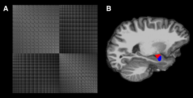
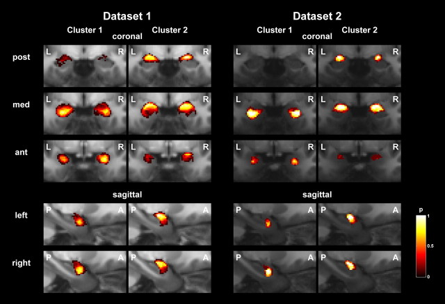
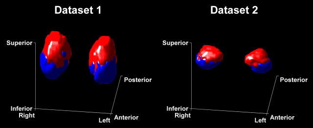
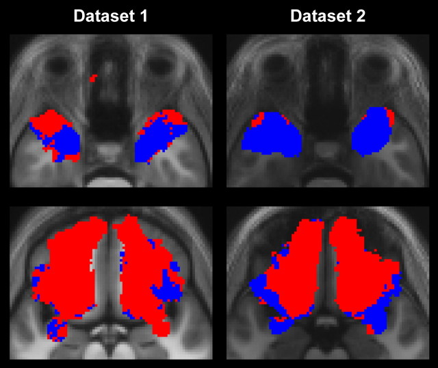
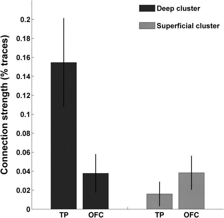
Despite a homogenous macroscopic appearance on magnetic resonance images, subregions of the amygdala express distinct functional profiles as well as corresponding differences in connectivity. In particular, histological analysis shows stronger connections for superficial (i.e., centromedial and cortical), compared with deep (i.e., basolateral and other), amygdala nuclei to lateral orbitofrontal cortex and stronger connections of deep compared with superficial, nuclei to polymodal areas in the temporal pole. Here, we use diffusion weighted imaging with probabilistic tractography to investigate these connections in humans. We use a data-driven approach to segment the amygdala into two subregions using k-means clustering. The identified subregions are spatially contiguous and their location corresponds to deep and superficial nuclear groups. Quantification of the connection strength between these amygdala clusters and individual target regions corresponds to qualitative histological findings in non-human primates, indicating such findings can be extrapolated to humans. We propose that connectivity profiles provide a potentially powerful approach for in vivo amygdala parcellation and can serve as a guide in studies that exploit functional and anatomical neuroimaging.
Figures
References
-
- Adolphs R, Spezio M. Role of the amygdala in processing visual social stimuli. Prog Brain Res. 2006;156:363–378. - PubMed
-
- Andersson JL, Skare S, Ashburner J. How to correct susceptibility distortions in spin-echo echo-planar images: application to diffusion tensor imaging. Neuroimage. 2003;20:870–888. - PubMed
-
- Ashburner J. A fast diffeomorphic image registration algorithm. Neuroimage. 2007;38:95–113. - PubMed
-
- Basser PJ, Mattiello J, LeBihan D. Estimation of the effective self-diffusion tensor from the NMR spin echo. J Magn Reson B. 1994a;103:247–254. - PubMed
Publication types
MeSH terms
Grants and funding
LinkOut - more resources
Full Text Sources