. 2011 Jan 12;31(2):669-77.
doi: 10.1523/JNEUROSCI.3659-10.2011.

EMMPRIN: a novel regulator of leukocyte transmigration into the CNS in multiple sclerosis and experimental autoimmune encephalomyelitis

Affiliations

EMMPRIN: a novel regulator of leukocyte transmigration into the CNS in multiple sclerosis and experimental autoimmune encephalomyelitis

Smriti M Agrawal et al. J Neurosci. .

Abstract

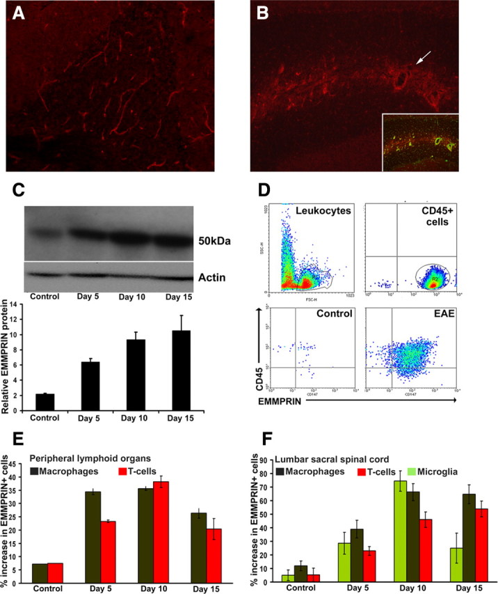
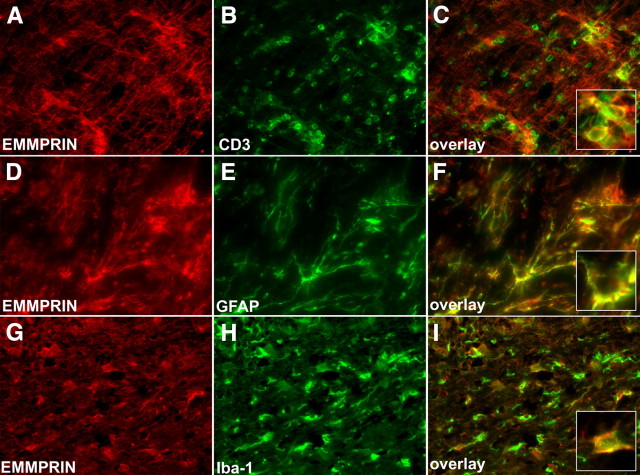
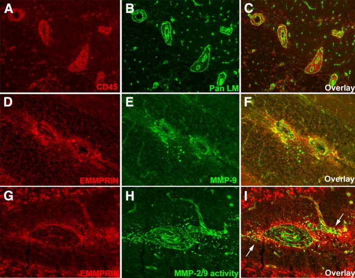
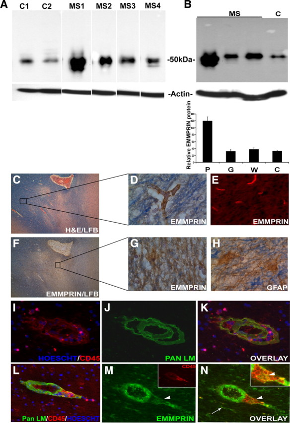
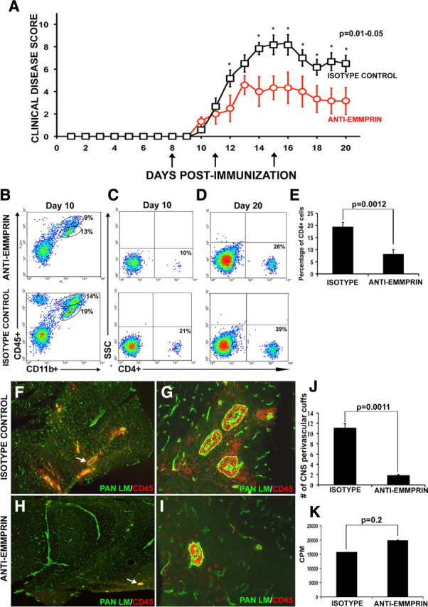
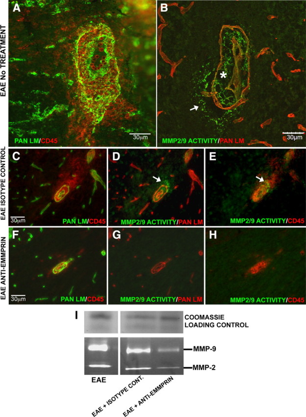
Extracellular matrix metalloproteinase inducer (EMMPRIN, CD147) is a member of the Ig superfamily, with various physiological roles including the induction of matrix metalloproteinases (MMPs), leukocyte activation, and tumor progression. In this study, we illustrate a novel involvement of EMMPRIN in multiple sclerosis (MS) and its animal model, experimental autoimmune encephalomyelitis (EAE). We found EMMPRIN levels to be upregulated on peripheral leukocytes before onset of EAE clinical signs and on infiltrating leukocytes and resident cells within the CNS in symptomatic mice. In EAE brain sections, EMMPRIN expression was localized with MMP-9 protein and activity. The increased EMMPRIN level was also characteristic of brain samples from MS subjects, particularly in plaque-containing areas. To evaluate the implications of elevated EMMPRIN levels, we treated EAE mice with an EMMPRIN function-blocking antibody and found reduced EAE clinical severity accompanied by decreased CNS parenchymal infiltration of leukocytes. Amelioration of EAE clinical signs by the anti-EMMPRIN antibody was critically dependent on its administration around the period of onset of clinical signs, which is typically associated with significant influx of leukocytes into the CNS. Moreover, the reduction in disease severity in anti-EMMPRIN-treated mice was associated with diminished MMP proteolytic activity at the glia limitans, the final barrier before parenchymal infiltration of leukocytes. Together, our results are the first to emphasize a role for EMMPRIN in MS and EAE, whereby EMMPRIN regulates leukocyte trafficking through increasing MMP activity. These results identify EMMPRIN as a novel therapeutic target in MS.


Figures

Figure 1.
Figure 1.
EMMPRIN expression is upregulated in EAE. Immunofluorescence staining (A, B), Western blot analysis (C), and flow cytometry (D–F) revealed higher EMMPRIN protein expression in mice immunized for EAE compared with naive controls. A, B, EMMPRIN expression was restricted to blood vessels in control cerebellum (A; 100× original magnification) and dramatically upregulated in EAE around inflamed blood vessels [B; 200× original magnification; the inset is double-immunofluorescence staining for pan laminin in green (vessels) and EMMPRIN in red] and also on other cellular profiles not associated with vessels. C, EMMPRIN, detected at 50 kDa in mouse spinal cord, increased (mean ± SEM; n = 3) in mice immunized for EAE at the presymptomatic stage (day 5), at the time of appearance of clinical signs (day 10), and at the peak clinical severity (day 15) compared with naive controls. Actin was used as a loading control. D, FACS analyses gating for CD45+ cells found increased numbers of EMMPRIN+ cells in lumbar sacral spinal cord isolates from EAE mice compared with controls. E, F, A time-point analysis on CD45+ gated cells from peripheral lymphoid organ (E) and lumbar sacral spinal cord (F) revealed increase in EMMPRIN in the T-cell (CD3+, red), macrophage (CD45highCD11bhigh, brown), and microglia (CD45low, CD11bhigh, green) populations with EAE progression. Data for fluorescence-activated cell sorting are mean ± SEM. pooled from nine mice each.
Figure 2.
Figure 2.
EMMPRIN is expressed by both CNS resident and infiltrating cells in EAE. At peak EAE disease (day 15), various cell types in the spinal cord were examined for EMMPRIN expression. A–C, Using immunofluorescence staining, we show that a large number of CD3+ infiltrating T-cells are also EMMPRIN positive; a high-magnification inset in C shows CD3 colocalization with EMMPRIN. D–F, GFAP+ astrocytes also expressed high amounts of EMMPRIN protein. G–I, Finally, Iba-1-positive macrophage/microglial cells stained positive for EMMPRIN. Pictures are representatives from similar data from nine mice each. All micrographs are 200× original magnification.
Figure 3.
Figure 3.
In EAE, EMMPRIN staining around inflammatory cuffs coincide with MMP-9 expression and MMP-2/9 activity. A–C, In the white matter of cerebellum of mice with peak EAE clinical severity, perivascular inflammatory cuffs typified by CD45+ leukocytes (red; A, C) sandwiched between the two laminin-containing (Pan LM; green; B, C) basement membranes of vessels are common (200× original magnification). D–F, High EMMPRIN levels (red; D, F) are found in and around the inflammatory cuffs coincident with high levels of MMP-9 protein (green; E, F) (200× original magnification). G–I, Similarly, EMMPRIN staining (red; G, I) colocalizes (I, arrows) with MMP-2/9 activity detected by in situ zymography (green; H, I) at sites of perivascular cuffs (400× magnification); in situ zymography cannot differentiate between the activity of MMP-2 and -9.
Figure 4.
Figure 4.
High EMMPRIN levels detected in MS plaques. A, Western blots revealed high EMMPRIN levels (50 kDa) in CNS samples from individual MS patients (MS1–2, with active lesions; MS3–4, inactive lesions) versus healthy donors (C1 and C2). Results are obtained from the same blot, and the white lines are attributable to irrelevant samples that have been spliced out. Similar levels of actin as the loading control in all samples are indicated. Samples C2 and MS4 have extra bands above and below the EMMPRIN band, respectively. The extra band in C2 is observed in a blot stained with the secondary antibody only (data not shown) and likely represents nonspecific binding. The lower bands in sample MS4 were not detected with the secondary antibody only and may be lower-molecular-weight glycosylated forms of EMMPRIN. B, Among the MS cases (where an example from one MS case is displayed), EMMPRIN was highly expressed in the MS plaque (P) compared to adjacent normal appearing white (W) and gray (G) matter. C, Hematoxylin and eosin with luxol fast blue staining of an MS plaque-containing specimen (40× original magnification) was used to examine EMMPRIN expression in both plaque and adjacent nonaffected white matter. D, E, G, Both immunohistochemistry (D) using 3,3′-diaminobenzidine tetrahydrochloride (DAB) and immunofluorescence staining (E) revealed EMMPRIN staining in normal-appearing areas to be restricted to blood vessels, whereas in plaques, EMMPRIN immunoreactivity was widespread (G). H, Immunohistochemistry for GFAP-positive astrocytes showed a similar pattern of expression as EMMPRIN. I–K, Immunofluorescence staining of an active MS plaque revealed the presence of inflammatory cuffs marked with pan laminin (Pan LM) staining for basement membranes surrounding the blood vessel and CD45+ leukocyte accumulation and infiltration into the CNS parenchyma. L–N, EMMPRIN, found to be predominantly expressed on endothelial cells of the inflammatory cuffs with this particular antibody, also colocalized with infiltrating CD45+ cells (M, inset; N, inset, arrowhead), and CNS resident cells (N, arrow). Hoescht dye was used to stain the nucleus of cells blue (I, K, L).
Figure 5.
Figure 5.
Treatment with anti-EMMPRIN function-blocking antibody attenuates EAE disease severity. A, Mice immunized for EAE were treated with an anti-EMMPRIN function-blocking antibody at days 8, 11, and 15 (arrows) after MOG immunization, and EAE disease severity was found to be significantly reduced compared with mice treated with an isotype antibody control. The data points that are statistically different are indicated (*p = 0.01–0.05, Mann–Whitney U test). Results are mean ± SEM. from six mice each; this was reproduced in another experiment. B–D, Using fluorescence-activated cell sorting, the percentage of CD45+CD11b+ macrophage/microglia (B; the ellipse with the higher CD45 represents macrophage, whereas the lower ellipse depicts microglia) and CD4+ T-cells at days 10 (C) and 20 (D) in the CNS were observed to be lower in mice treated with anti-EMMPRIN compared with those treated with isotype control. E, Percentage of CD4+ T-cells was lower in the CNS of EAE mice treated with anti-EMMPRIN compared with treatment with isotype control. F–J, Immunofluorescence staining for pan laminin (green) and CD45+ (red) cells of mouse cerebellum reveal a significantly higher number of perivascular cuffs (arrows) and CNS parenchymal infiltrates in isotype-treated [40× (F) and 200× (G) original magnification, respectively] animals compared with those treated with anti-EMMPRIN [40× (H) and 200× (I) original magnification, respectively]; the quantified results of the total number of perivascular cuffs observed in both groups is displayed in J. Results are representative from six mice in each group. K, Importantly, the recall response of T-cells to MOG35–55 was not significantly different between the anti-EMMPRIN- and isotype control-treated groups. CPM, Counts per minute in a proliferation assay.
Figure 6.
Figure 6.
Anti-EMMPRIN antibody treatment reduces MMP activity in the CNS. A–C, Inflamed vessels with numerous CD45+ cells both within the perivascular cuff and invading the CNS parenchyma (B; asterisk and arrow, respectively) were observed in cerebellum cryosections of EAE mice with no treatment or isotype control antibody treatment (A, C), double-immunofluorescence stained for pan laminin (green) and CD45+ (red). B, D, E, Immunofluorescence staining for pan laminin (red) coupled with in situ zymography (green) showed MMP activity corresponding to the sites where leukocytes infiltrate the glia limitans to enter the CNS parenchyma. F, In EAE mice treated with anti-EMMPRIN antibody, double-immunofluorescence staining showed CD45+ cells to be restricted to the perivascular cuff, with very little to no infiltration into the CNS parenchyma. G–I, Immunofluorescence coupled with in situ zymography for MMP activity and gelatin gel zymography showed a reduction in MMP-2 and MMP-9 activity and levels with anti-EMMPRIN treatment. Coomassie blue was used to stain a nongelatin gel to verify equal sample loading on gel zymograms (I). In all cases, results are representatives from two independent experiments with at least three animals per treatment in each group.

References

    1. Agrawal S, Anderson P, Durbeej M, van Rooijen N, Ivars F, Opdenakker G, Sorokin L. Dystroglycan is selectively cleaved at the parenchymal basement membrane at sites of leukocyte extravasation in experimental autoimmune encephalomyelitis. J Exp Med. 2006;203:1007–1019. - PMC - PubMed
    1. Anthony DC, Ferguson B, Matyzak MK, Miller KM, Esiri MM, Perry VH. Differential matrix metalloproteinase expression in cases of multiple sclerosis and stroke. Neuropathol Appl Neurobiol. 1997;23:406–415. - PubMed
    1. Berditchevski F, Chang S, Bodorova J, Hemler ME. Generation of monoclonal antibodies to integrin-associated proteins. Evidence that alpha3beta1 complexes with EMMPRIN/basigin/OX47/M6. J Biol Chem. 1997;272:29174–29180. - PubMed
    1. Bernal F, Elias B, Hartung HP, Kieseier BC. Regulation of matrix metalloproteinases and their inhibitors by interferon-beta: a longitudinal study in multiple sclerosis patients. Mult Scler. 2009;15:721–727. - PubMed
    1. Biswas C, Zhang Y, DeCastro R, Guo H, Nakamura T, Kataoka H, Nabeshima K. The human tumor cell-derived collagenase stimulatory factor (renamed EMMPRIN) is a member of the immunoglobulin superfamily. Cancer Res. 1995;55:434–439. - PubMed

Publication types