Comparative Study
. 2011 May;84(5):976-85.
doi: 10.1095/biolreprod.110.087460. Epub 2011 Jan 12.

ATP synthesis, mitochondrial function, and steroid biosynthesis in rodent primary and tumor Leydig cells

Affiliations
Comparative Study

ATP synthesis, mitochondrial function, and steroid biosynthesis in rodent primary and tumor Leydig cells

Andrew S Midzak et al. Biol Reprod. 2011 May.

Abstract

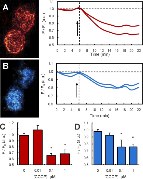
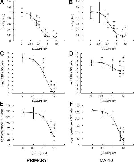
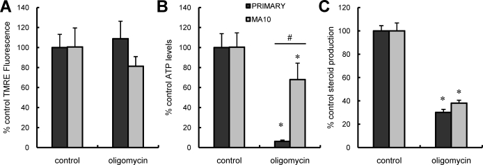
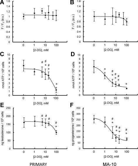
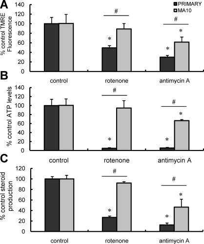
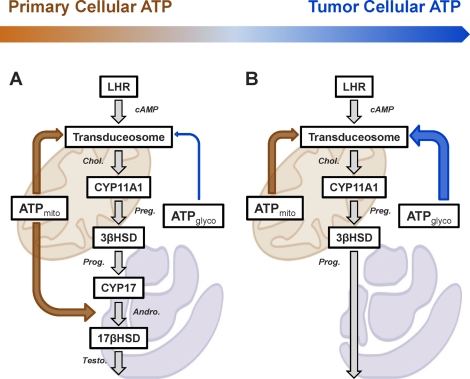
Previous studies in MA-10 tumor Leydig cells demonstrated that disruption of the mitochondrial electron-transport chain (ETC), membrane potential (ΔΨ(m)), or ATP synthesis independently inhibited steroidogenesis. In contrast, studies of primary Leydig cells indicated that the ETC, ΔΨ(m), and ATP synthesis cooperatively affected steroidogenesis. These results suggest significant differences between the two systems and call into question the extent to which results from tumor Leydig cells relate to primary cells. Thus, to further understand the similarities and differences between the two systems as well as the impact of ATP disruption on steroidogenesis, we performed comparative studies of MA-10 and primary Leydig cells under similar conditions of mitochondrial disruption. We show that mitochondrial ATP synthesis is critical for steroidogenesis in both primary and tumor Leydig cells. However, in striking contrast to primary cells, perturbation of ΔΨ(m) in MA-10 cells did not substantially decrease cellular ATP content, a perplexing finding because ΔΨ(m) powers the mitochondrial ATP synthase. Further studies revealed that a significant proportion of cellular ATP in MA-10 cells derives from glycolysis. In contrast, primary cells appear to be almost completely dependent on mitochondrial respiration for their energy provision. Inhibitor studies also suggested that the MA-10 ETC is impaired. This work underscores the importance of mitochondrial ATP for hormone-stimulated steroid production in both MA-10 and primary Leydig cells while indicating that caution must be exercised in extrapolating data from tumor cells to primary tissue.


Figures

FIG. 1.
FIG. 1.
Two-photon microscopy (TPM) of mitochondrial ΔΨm and cellular NAD(P)H levels in LH-stimulated and in LH- and CCCP-stimulated primary Leydig cells. A) TPM image of TMRM fluorescence from two Leydig cells and corresponding time traces of TMRM fluorescence in the same cells exposed to LH (100 ng/ml) and to LH plus 1 μM CCCP (arrow). B) TPM image of NAD(P)H autofluorescence from the same Leydig cells as in A and corresponding time traces of NAD(P)H autofluorescence in the same cells exposed to LH (100 ng/ml) and to LH and 1 μM CCCP (arrow). C) TPM readings (mean ± SEM) of TMRM fluorescence of Leydig cells exposed to increasing concentrations of CCCP (0–1 μM) (summary of two experiments, n = 20 cells). D) TPM readings (mean ± SEM) of NAD(P)H autofluorescence of Leydig cells exposed to increasing concentrations of CCCP (0–1 μM) (summary of two experiments, n = 20 cells). *P < 0.001 vs. cells incubated without CCCP.
FIG. 2.
FIG. 2.
Effect of CCCP on LH-stimulated primary and MA-10 tumor Leydig cell mitochondrial ΔΨm, intracellular ATP content, and steroid production. A and B) Fluorescence plate reader assay of ΔΨm in primary (A) and MA-10 tumor (B) Leydig cultures incubated with increasing doses of CCCP (0–10 μM) in the presence of maximally stimulating LH (100 ng/ml) for 15 min. C and D) ATP levels of primary (C) and MA-10 tumor (D) Leydig cultures incubated with increasing doses of CCCP (0–10 μM) in the presence of maximally stimulating LH (100 ng/ml) for 2 h. E and F) Steroid production by primary (E) and MA-10 tumor (F) Leydig cultures were incubated with increasing doses of CCCP (0–10 μM) in the presence of maximally stimulating LH (100 ng/ml) for 2 h. Points shown represent the mean ± SEM of three to four experiments, with three replicates per experiment. *P < 0.01 vs. cells incubated without CCCP, #P < 0.05 vs. corresponding primary or MA-10 cells.
FIG. 3.
FIG. 3.
Effect of oligomycin on LH-stimulated mitochondrial ΔΨm, intracellular ATP content, and steroid production in primary and MA-10 tumor Leydig cells. Black bars correspond to controls; gray bars correspond to oligomycin treatment. Data are presented as a percentage of control values. A) ΔΨm in primary and MA-10 tumor Leydig cultures incubated with oligomycin (1 μg/ml) in the presence of maximally stimulating LH (100 ng/ml) for 15 min. B) ATP levels of primary and MA-10 tumor Leydig cultures incubated with oligomycin (1 μg/ml) in the presence of maximally stimulating LH (100 ng/ml) for 2 h. C) Steroid production by primary and MA-10 tumor Leydig cultures incubated with oligomycin (1 μg/ml) in the presence of maximally stimulating LH (100 ng/ml) for 2 h. Values shown represent the mean ± SEM of three experiments, with three replicates per experiment. *P < 0.001 vs. cells incubated without oligomycin, #P < 0.05 vs. corresponding primary or MA-10 cells.
FIG. 4.
FIG. 4.
Effect of 2-DG on LH-stimulated primary and MA-10 tumor Leydig cell mitochondrial ΔΨm, intracellular ATP content, and steroid production. A and B) ΔΨm in primary (A) and MA-10 tumor (B) Leydig cultures incubated with increasing doses of 2-DG (0–100 mM) in the presence of maximally stimulating LH (100 ng/ml) for 15 min. C and D) ATP levels in primary (C) and MA-10 tumor (D) Leydig cultures incubated with increasing doses of 2-DG (0–100 mM) in the presence of maximally stimulating LH (100 ng/ml) for 2 h. E and F) Steroid production by primary (E) and MA-10 tumor (F) Leydig cultures incubated with increasing doses of 2-DG (0–100 mM) in the presence of maximally stimulating LH (100 ng/ml) for 2 h. Points shown represent the mean ± SEM of three experiments, with three replicates per experiment. *P < 0.01 vs. cells incubated with CCCP, #P < 0.05 vs. corresponding primary or MA-10 cells.
FIG. 5.
FIG. 5.
Effect of ETC inhibition on LH-stimulated mitochondrial ΔΨm, intracellular ATP content, and steroid production in primary and MA-10 tumor Leydig cells. Black bars correspond to controls; gray bars correspond to rotenone or antimycin A treatment. Data are presented as a percentage of control values. A) ΔΨm in primary and MA-10 tumor Leydig cultures incubated with rotenone (0.1 μM) or antimycin A (1 μM) in the presence of maximally stimulating LH (100 ng/ml) for 15 min. B) ATP levels in primary and MA-10 tumor Leydig cultures incubated with rotenone (0.1 μM) or antimycin A (1 μM) in the presence of maximally stimulating LH (100 ng/ml) for 2 h. C) Steroid production by primary and MA-10 tumor Leydig cells incubated with rotenone (0.1 μM) or antimycin A (1 μM) in the presence of maximally stimulating LH (100 ng/ml) for 2 h. Points shown represent the mean ± SEM of three experiments, with three replicates per experiment. *P < 0.001 vs. cells incubated without ETC inhibitors, #P < 0.05 vs. corresponding primary or MA-10 cells.
FIG. 6.
FIG. 6.
Effect of mitochondrial disruption and glycolytic inhibition on metabolic flux through steroidogenic pathway in primary Leydig cells. A) ATP levels in cells incubated with 1 μg/ml of oligomycin or 5 μM CCCP in the presence of either LH (100 ng/ml), dbcAMP (1 mM), HC (20 μM), P5 (10 μM), P4 (10 μM), or androstenedione (5 μM) for 2 h. B) Testosterone production in cells treated as in A. After 2 h, medium was collected and testosterone levels assessed by RIA. Values are presented as the percentage of testosterone production without inhibitor. C) ATP levels in cells incubated with 100 mM 2-DG in the presence of either LH (100 ng/ml), dbcAMP (1 mM), HC (20 μM), P5 (10 μM), P4 (10 μM), or androstenedione (5 μM) for 2 h. D) Testosterone production in cells treated as in C. After 2 h, medium was collected and testosterone levels assessed by RIA. Values are presented as the percent of testosterone production without inhibitor. Lowercase letters designate groups that are statistically significant from each other (P < 0.05).
FIG. 7.
FIG. 7.
Model of Leydig cell ATP utilization. Hormone-responsive primary (A) and tumor (B) Leydig cells critically utilize mitochondrial ATP for mitochondrial cholesterol transport (upper orange arrow). However, the tumorigenic transition has resulted in a larger glycolytic contribution to cellular ATP (gradient arrow above). Consequently, tumor Leydig cells utilize a greater proportion of glycolytic ATP (B; blue arrow) for cholesterol transport than do primary cells (A; blue arrow). Primary cells also utilize mitochondria-derived ATP for enzymatic reactions taking place in the ER (A; lower orange arrow), which are missing in tumor Leydig cells (B).

References

    1. Puett D, Li Y, DeMars G, Angelova K, Fanelli F. A functional transmembrane complex: the luteinizing hormone receptor with bound ligand and G protein. Mol Cell Endocrinol 2007; 260–262: 126 136 - PMC - PubMed
    1. Miller WL. Steroidogenic acute regulatory protein (StAR), a novel mitochondrial cholesterol transporter. Biochim Biophys Acta 2007; 1771: 663 676 - PubMed
    1. Papadopoulos V, Baraldi M, Guilarte TR, Knudsen TB, Lacapère JJ, Lindemann P, Norenberg MD, Nutt D, Weizman A, Zhang MR, Gavish M. Translocator protein (18 kDa): new nomenclature for the peripheral-type benzodiazepine receptor based on its structure and molecular function. Trends Pharmacol Sci 2006; 27: 402 409 - PubMed
    1. Rone MB, Fan J, Papadopoulos V. Cholesterol transport in steroid biosynthesis: role of protein-protein interactions and implications in disease states. Biochim Biophys Acta 2009; 1791: 646 658 - PMC - PubMed
    1. Liu J, Rone MB, Papadopoulos V. Protein-protein interactions mediate mitochondrial cholesterol transport and steroid biosynthesis. J Biol Chem 2006; 281: 38879 38893 - PubMed

Publication types

MeSH terms