. 2011 Apr;85(7):3120-7.
doi: 10.1128/JVI.02004-10. Epub 2011 Jan 12.

Regulation of the human papillomavirus type 18 E6/E6AP ubiquitin ligase complex by the HECT domain-containing protein EDD

Affiliations

Regulation of the human papillomavirus type 18 E6/E6AP ubiquitin ligase complex by the HECT domain-containing protein EDD

Vjekoslav Tomaic et al. J Virol. 2011 Apr.

Abstract

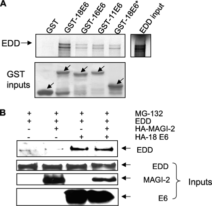
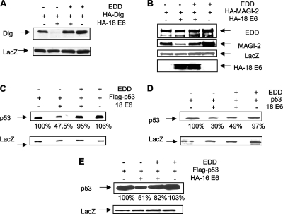
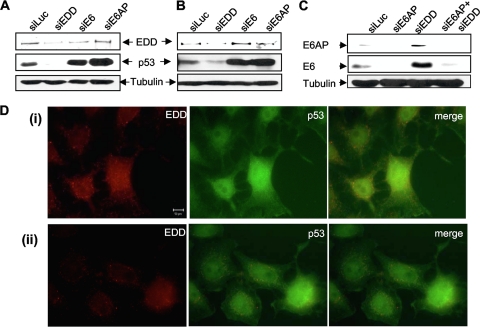
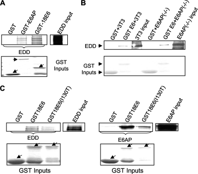
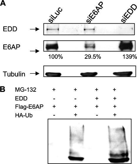
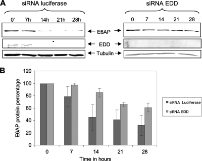
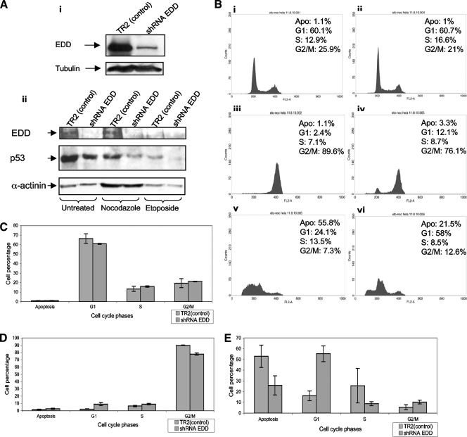
Human papillomavirus (HPV) E6 oncoproteins target many cellular proteins for ubiquitin-mediated proteasomal degradation. In the case of p53, this is mediated principally by the E6AP ubiquitin ligase. Several studies have reported that E6 can target certain of its substrates in an apparently E6AP-independent fashion and that several of these substrates vary in the degree to which they are degraded by E6 at different stages of malignancy. To more fully understand the regulation of the E6AP/E6 proteolytic targeting complex, we performed a mass spectroscopic analysis of HPV type 18 (HPV-18) E6 protein complexes and identified the HECT domain-containing ubiquitin ligase EDD as a new HPV-18 E6 binding partner. We show that EDD can interact independently with both E6 and E6AP. Furthermore, EDD appears to regulate E6AP expression levels independently of E6, and loss of EDD stimulates the proteolytic activity of the E6/E6AP complex. Conversely, higher levels of EDD expression protect a number of substrates from E6-induced degradation, partly as a consequence of lower levels of E6 and E6AP expression. Intriguingly, reduction in EDD expression levels in HPV-18-positive HeLa cells enhances cell resistance to apoptotic and growth arrest stimuli. These studies suggest that changes in the levels of EDD expression during different stages of the viral life cycle or during malignancy could have a profound effect upon the ability of E6 to target various substrates for proteolytic degradation and thereby directly influence the development of HPV-induced malignancy.


Figures

FIG. 1.
FIG. 1.
HPV-18 E6 protein binds to EDD in vitro and in vivo. (A) Radiolabeled in vitro translated EDD was incubated with GST, GST-18E6, GST-16E6, GST-11E6, and GST-18E6*. Bound proteins were assessed by autoradiography, and the input GST fusion proteins were visualized with Coomassie staining (lower panel). Input EDD (10%) is shown. (B) 293 cells were transfected with HA-tagged HPV-18 E6 (HA-18E6), EDD1, or HA-tagged MAGI-2, as indicated. After 24 h cells were incubated for 3 h with MG132 before being harvested, and cell extracts were immunoprecipitated with anti-HA antibody. Coprecipitating EDD was detected by Western blotting with anti-EDD antibody. The three lower panels show the protein inputs of EDD, MAGI-2, and HPV-18 E6.
FIG. 2.
FIG. 2.
EDD inhibits HPV-18 E6 degradation of Dlg, MAGI-2, and p53. 293 cells (A, B, C, and E) or p53 null H1299 cells (D) were transfected with HA-tagged Dlg, HA-MAGI-2, EDD1, and HPV-18 E6 (A, B, C, and D) or HPV-16 E6 (E), alone or in combination. After 24 h cells were harvested, and residual Dlg (A), MAGI-2 (B), and p53 (C, D, and E) were detected by Western blot analysis using either anti-HA antibody (A, B, and C) or anti-p53 antibody (D) or by anti-Flag where p53 was Flag tagged (C and E). The expression of ß-galactosidase (LacZ) was used as a control of transfection efficiency and loading (lower panels), and the percentage of p53 remaining in each track in panels C, D, and E is also shown.
FIG. 3.
FIG. 3.
EDD knockdown enhances E6 activity. HeLa cells (A and C) and CaSKi cells (B) were transfected with siRNAs directed against luciferase (siLuc), EDD (siEDD), E6AP (siE6AP), or HPV-16 E6/E7 or HPV-18 E6/E7 (siE6), alone or in combination with siE6AP and siEDD. After 72 h cells were harvested, and the levels of EDD, p53, E6, and the tubulin loading control were detected by Western blotting. HeLa cells (D) were fixed and probed with goat anti-EDD and rabbit anti-p53 antibodies, followed by rhodamine-conjugated donkey anti-goat (red for EDD) and fluorescein isothiocyanate-conjugated donkey anti-rabbit (green for p53) antibodies. Two different fields are shown (i and ii).
FIG. 4.
FIG. 4.
E6 does not require E6AP to bind EDD. (A) In vitro translated EDD was incubated with GST, GST-E6AP, and GST-18E6. Bound proteins were assessed by autoradiography, and the input GST fusion proteins were visualized with Coomassie staining (lower panel). Input EDD (10%) is shown. (B) Extracts of E6AP−/− and 3T3 cells were incubated with GST and GST-18E6 fusion proteins for 2 h at 4°C. Bound proteins were assessed by Western blotting using EDD antibody. The EDD inputs from the cells are also shown. (C) In vitro translated EDD and E6AP were incubated with GST, GST-18E6, and GST-18E6 (I130T). Bound proteins were assessed by autoradiography, and the input GST fusion proteins were visualized with Coomassie staining (lower panel). GST-18 E6 (I130T) binding to EDD is 21% lower than that of wild-type GST-18E6, while GST-18E6 (I130T) binding to E6AP is 77% lower than that of wild-type GST-18E6. Arrows indicate GST fusion proteins.
FIG. 5.
FIG. 5.
siEDD increases E6AP levels in HPV-negative cells. (A) HT1080 cells were transfected with siLuc, siE6AP, or siEDD. After 72 h cells were harvested, and levels of EDD, E6AP, and tubulin were determined by Western blotting. The numbers show the percentages of E6AP remaining. (B) 293 cells were cotransfected with EDD, Flag-E6AP, and HA-ubiquitin (HA-Ub), and after 24 h cells were treated for 3 h with the proteasome inhibitor MG132. The cells were then harvested, and complexes were immunoprecipitated with anti-HA-conjugated agarose beads. Complexes were then analyzed by Western blotting for E6AP using anti-Flag antibodies. Note the increased levels of mono- and polyubiquitinated forms of E6AP in the EDD-expressing cells.
FIG. 6.
FIG. 6.
E6AP protein turnover is regulated by EDD. (A) HeLa cells were transfected with siLuc or siEDD. At 72 h posttransfection cells were treated with cycloheximide at different time points (0, 7 h, 14 h, 21 h, and 28 h). E6AP, EDD, and tubulin levels were detected by Western blotting. (B) Collated results from three independent experiments to measure residual E6AP protein levels, with band intensities determined using the OptiQuant quantification program. E6AP levels were normalized to 100% at time zero. Standard deviations are also shown.
FIG. 7.
FIG. 7.
EDD knockdown inhibits checkpoint-activated cell growth arrest and apoptosis. (A). HeLa cells stably transfected with shRNA EDD and with nonspecific shRNA TR2 (control) were harvested in SDS sample buffer, and residual EDD levels were assessed by Western blotting (i). The lower panel (ii) shows Western blots of EDD and p53 in the control and EDD-ablated cells growing asynchronously and postexposure to nocodazole and etoposide. (B) FACS analysis showing the cell cycle profiles of asynchronously growing cells (i and ii) and following nocodazole (iii and iv) and etoposide treatments (v and vi). Control cells are shown in panels i, iii, and v, with EDD knockdown cells in panels ii, iv and vi. Apo, apoptosis. (C, D, and E) Percentages of cells in each phase of the ell cycle in asynchronously growing cells (C) and following treatment with nocodazole (D) and etoposide (E) from at least three separate experiments; the standard deviations are also shown.

References

    1. Banks, L., S. C. Barnett, and T. Crook. 1990. HPV-16 E7 functions at the G1 to S phase transition in the cell cycle. Oncogene 5:833-837. - PubMed
    1. Beer-Romero, P., S. Glass, and M. Rolfe. 1997. Antisense targeting of E6AP elevates p53 in HPV-infected cells but not in normal cells. Oncogene 14:595-602. - PubMed
    1. Boyer, S. N., D. E. Wazer, and V. Band. 1996. E7 protein of human papilloma virus-16 induces degradation of retinoblastoma protein through the ubiquitin-proteasome pathway. Cancer Res. 56:4620-4624. - PubMed
    1. Brimer, N., C. Lyons, and S. B. Vande Pol. 2007. Association of E6AP (UBE3A) with human papillomavirus type 11 E6 protein. Virology 358:303-310. - PMC - PubMed
    1. Callaghan, M. J., et al. 1998. Identification of a human HECT family protein with homology to the Drosophila tumor suppressor gene hyperplastic discs. Oncogene 17:3479-3491. - PubMed

Publication types

MeSH terms

Substances