Review
. 2011 Apr;69(4):271-8.
doi: 10.1203/PDR.0b013e31820efbcf.

Adverse and protective influences of adenosine on the newborn and embryo: implications for preterm white matter injury and embryo protection

Affiliations
Review

Adverse and protective influences of adenosine on the newborn and embryo: implications for preterm white matter injury and embryo protection

Scott A Rivkees et al. Pediatr Res. 2011 Apr.

Abstract

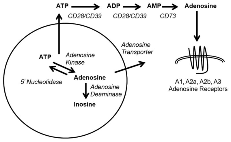
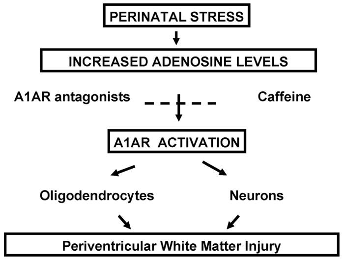
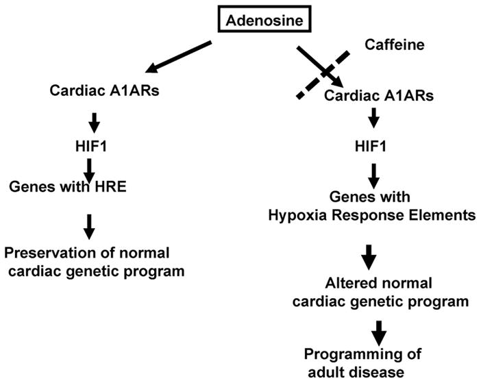
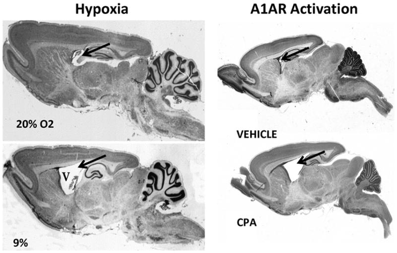
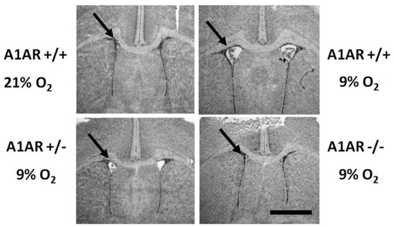
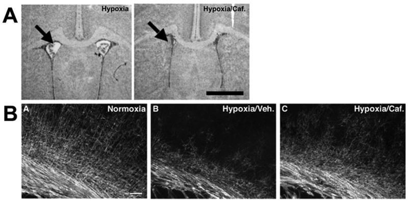
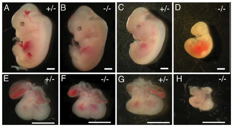
Few signaling molecules have the potential to influence the developing mammal as the nucleoside adenosine. Adenosine levels increase rapidly with tissue hypoxia and inflammation. Adenosine antagonists include the methylxanthines caffeine and theophylline. The receptors that transduce adenosine action are the A1, A2a, A2b, and A3 adenosine receptors (ARs). In the postnatal period, A1AR activation may contribute to white matter injury in the preterm infant by altering oligodendrocyte (OL) development. In models of perinatal brain injury, caffeine is neuroprotective against periventricular white matter injury (PWMI) and hypoxic-ischemic encephalopathy (HIE). Supporting the notion that blockade of adenosine action is of benefit in the premature infant, caffeine reduces the incidence of bronchopulmonary dysplasia and CP in clinical studies. In comparison with the adverse effects on the postnatal brain, adenosine acts via A1ARs to play an essential role in protecting the embryo from hypoxia. Embryo protective effects are blocked by caffeine, and caffeine intake during early pregnancy increases the risk of miscarriage and fetal growth retardation. Adenosine and adenosine antagonists play important modulatory roles during mammalian development. The protective and deleterious effects of adenosine depend on the time of exposure and target sites of action.


Figures

Figure 1
Figure 1. Adenosine action
Adenosine is generated at intra- and extracellular sites from the breakdown of adenosine triphosphate. Extracellular adenosine binds to G-protein-coupled P1 purine receptors, A1, A2a, A2b and A3. A1 and A3 receptors are Gi/o-coupled. A2a and A2b receptors are Gs-coupled and increase intracellular cyclic AMP levels.
Figure 2
Figure 2. A1AR mRNA and binding site expression in rat brain
Sense (A) and antisense (B) images generated from in situ hybridization studies are shown. (C) Specific [3H]DPCPX labeling by receptor autoradiography. Specific labeling appears as white. Note the mismatch between patterns of mRNA and receptor labeling. mRNA is expressed over cells in the granular layer of the dentate gyrus (dg) and over cells in the pyramidal layer (py) of Ammon’s horn. In contrast [3H]DPCPX labeling of A1ARs is greatest in the dentate gyrus molecular layer (dm), the polymorphic (po) and molecular layers (am). Scale bar = 25 mm. Reprinted from Swanson TH et al. J Comp Neurol 363:517–531; Copyright c 1995 Wiley-Liss, Inc., with permission.
Figure 3
Figure 3. A1 adenosine receptor (A1AR) activation induces periventricular white matter injury
Left panel: Neonatal rats reared in hypoxia manifest features of periventricular white matter injury including white matter loss and secondary ventriculomegaly (V). Reprinted from Ment LR et al. Brain Res Dev Brain Res 111:197–203; Copyright c 1998 Elsevier Science B.V., with permission. Right Panel: Neonatal rats treated with the A1 adenosine receptor (A1AR) agonist, N6-cylcopentyladenosine (CPA) manifest features of PWMI similar to that observed in hypoxia. Arrows depict location of ventricles.
Figure 4
Figure 4. Deletion of A1ARs protects against periventricular white matter injury
Hematoxylin-stained coronal sections from A1AR +/+, +/−, or −/− animals taken from the midstriatum of P14 mice exposed from P3 through P14 to either chronic sublethal hypoxia (9.5% O2) or room air. Ventricular enlargement was observed in +/+ and +/− mice exposed to hypoxia but not in −/− mice exposed to hypoxia and +/+ mice reared in normoxia. Scale bar: 1 mm. Reprinted from Turner CP et al. Proc Natl Acad Sci USA 100:11718–11722; Copyright c 2003 The National Academy of Sciences of the U.S.A., with permission.
Figure 5
Figure 5. Caffeine protects against periventricular white matter injury
(A) Representative coronal views of the lateral ventricles (arrow) for mice reared in hypoxia by dams drinking water (upper panel) or water containing caffeine (CAF; lower panel). Ventriculomegaly was not seen in mice reared in hypoxia by dams drinking water with caffeine. (B) Caffeine treatment during chronic sublethal hypoxia ameliorates reductions in cerebral myelination. Myelin basic protein (MBP) staining shows the typical myelination patterns of animals reared in normoxia (A) relative to those in chronic hypoxia treated with vehicle (Veh.; B) or with caffeine (Caf.; C). Scale bar: 1 mm. Reprinted from Back SA Ann Neurol 60:696–705; Copyright c 2006 American Neurological Association, with permission.
Figure 6
Figure 6. A1ARs protects against growth retardation in embryos
Hypoxia induces severe growth retardation in A1AR−/− embryos. Dams were exposed to 10% O2 from E8.5 to E12.5. C–R length was measured for normoxia and hypoxia-treated embryos at E12.5. Under normoxia conditions (A). A1AR+/− embryos were indistinguishable from A1AR−/− (B) embryos. Under hypoxic conditions, A1AR+/− embryos (C) were smaller then the normoxic controls (A), but A1AR−/− embryos (D) were significantly smaller than A1AR−/− or A1AR+/− normoxic embryos. A1AR−/− hypoxic hearts (H) were smaller than A1AR+/− normoxic (E), A1AR−/− normoxic (F), or A1AR+/− hypoxic hearts (G). Scale bars: 1 mm. Reprinted from Wendler CC et al. Proc Natl Acad Sci USA 104:9697–9702; Copyright c 2007 The National Academy of Sciences of the U.S.A., with permission.
Figure 7
Figure 7. Cardiac A1ARs protect against hypoxia
Embryos exposed to hypoxia for 2 days in utero from E8.5-10.5 were not growth retarded compared to room air controls, but embryos exposed to hypoxia in utero from E10-12.5 exhibited significant growth retardation. Embryos exposed to hypoxia for 3 days demonstrated an even greater amount of growth retardation compared to controls. Normoxic embryos both (A) Normox/Flox and (B) Normox/Cre displayed normal morphology and growth. The hypoxic embryos (C) Hypox/Flox and (D) Hypox/Cre were significantly growth retarded, however there was no difference between Hypox/Flox and Hypox/Cre embryos. Scale bar is 1 mm. Reprinted with authors’ permission from Wendler CC et al. BMC Dev Biol 10:57; Copyright c 2010 Wendler et al.
Figure 8
Figure 8. Hypoxia and caffeine treatment lead to reduced ventricular myocardial tissue
Embryos were exposed to hypoxia and caffeine from E8.5–E10.5. Compared to NorNS (A), ventricular myocardial area was decreased by 37.3% in the NorCf (B) group. Exposure to hypoxia caused a more substantial decrease in myocardial area, including 55.7% in the HyNS group (C) and 53.3% in the HyCf group (D5). V, ventricle, *, endocardial cushion. Scale bars = 100 μm. Reprinted from Wendler CC et al. FASEB J 23:1272–1278; Copyright c 2009 FASEB, with permission.
Figure 9
Figure 9. Caffeine blocks hypoxia-induced HIF1 protein accumulation in hypoxic embryos
Caffeine treatment inhibited HIF1 protein accumulation in hypoxic embryos. Dams were injected with normal saline or 20 mg/kg caffeine and then placed immediately in hypoxia (10% O2) or left in room air (21% O2). Embryos were collected after 6 hr, and whole embryo protein was isolated. Western blot analysis of HIF1 protein expression demonstrated a 40% reduction in stabilized HIF1 protein in caffeine-treated embryos exposed to hypoxia. β-Actin protein was examined on the same Western blots as a loading control. Reprinted from Wendler CC et al. FASEB J 23:1272– 1278; Copyright c 2009 FASEB, with permission.
Figure 10
Figure 10
Cartoon depicting contribution of adenosine to periventricular white matter injury.
Figure 11
Figure 11
Cartoon depicting contribution of adenosine to protecting the embryo against intrauterine stress and how this is disrupted by caffeine.

References

    1. Jacobson KA. Introduction to adenosine receptors as therapeutic targets. Handb Exp Pharmacol. 2009;193:1–24. - PMC - PubMed
    1. Rivkees SA, Zhao Z, Porter G, Turner C. Influences of adenosine on the fetus and newborn. Mol Genet Metab. 2001;74:160–171. - PubMed
    1. Eckle T, Koeppen M, Eltzschig HK. Role of extracellular adenosine in acute lung injury. Physiology (Bethesda) 2009;24:298–306. - PubMed
    1. Eltzschig HK, Weissmuller T, Mager A, Eckle T. Nucleotide metabolism and cell-cell interactions. Methods Mol Biol. 2006;341:73–87. - PubMed
    1. Eltzschig HK, Abdulla P, Hoffman E, Hamilton KE, Daniels D, Schonfeld C, Loffler M, Reyes G, Duszenko M, Karhausen J, Robinson A, Westerman KA, Coe IR, Colgan SP. HIF-1-dependent repression of equilibrative nucleoside transporter (ENT) in hypoxia. J Exp Med. 2005;202:1493–1505. - PMC - PubMed

Publication types

MeSH terms