

Comment
doi: 10.1038/469172a.
Cell signalling: Binding the receptor at both ends
- PMID: 21228868
- PMCID: PMC3804163
- DOI: 10.1038/469172a
Item in Clipboard
Comment
Cell signalling: Binding the receptor at both ends
Nature.
.
Abstract
G-protein-coupled receptors initiate a wide range of signalling pathways in cells. It seems that both a G protein and an agonist molecule must bind to the receptors to persistently activate them.
Figures
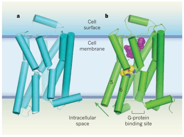
G-protein-coupled receptors (GPCRs) are a family of membrane-bound proteins. They
activate G proteins when bound by agonist molecules, but are inactive in the
presence of inverse agonists. a, The structure of the
β-adrenergic receptor β2AR — a GPCR — is
shown here in its inactive (R) state. The receptor is bound to the inverse agonist carazolol (not
shown). Cylinders indicate α-helices. b, Rasmussen
et al.
report the crystal structure of β2AR in complex with an
agonist (purple) and with an antibody fragment (not shown), both of which
stabilize the receptor’s active (R*) state. The antibody binds at the
G-protein binding site, and acts as a substitute for
β2AR’s G protein (Gs). The transition from
the R to the R* state, induced by agonist binding, probably results from a
contraction of the agonist binding site. The contraction promotes changes in the
packing of amino-acid residues (yellow/blue/red spheres)
between transmembrane helices 5 and 6. These changes coincide with rotation of
parts of these helices (green arrow). Together, the conformational shifts create
a binding site for the carboxy terminus of the G protein (or, in this case, the
antibody fragment). This crystal structure, along with structures and molecular simulations in two
other papers,, suggest that both a G protein and an agonist
must bind to GPCRs to stabilize the R* state.
Comment on
-
Structure of a nanobody-stabilized active state of the β(2) adrenoceptor.Nature. 2011 Jan 13;469(7329):175-80. doi: 10.1038/nature09648. Nature. 2011. PMID: 21228869 Free PMC article.
-
Structure and function of an irreversible agonist-β(2) adrenoceptor complex.Nature. 2011 Jan 13;469(7329):236-40. doi: 10.1038/nature09665. Nature. 2011. PMID: 21228876 Free PMC article.
-
The structural basis for agonist and partial agonist action on a β(1)-adrenergic receptor.Nature. 2011 Jan 13;469(7329):241-4. doi: 10.1038/nature09746. Nature. 2011. PMID: 21228877 Free PMC article.
References
-
- Pierce KL, Premont RT, Lefkowitz RJ. Nature Rev. Mol. Cell Biol. 2002;3:639–650. - PubMed
Publication types
MeSH terms
Substances
Grants and funding
LinkOut - more resources
Full Text Sources
