Comparative Study
. 2011 Jan 13;469(7329):241-4.
doi: 10.1038/nature09746.

The structural basis for agonist and partial agonist action on a β(1)-adrenergic receptor

Affiliations
Comparative Study

The structural basis for agonist and partial agonist action on a β(1)-adrenergic receptor

Tony Warne et al. Nature. .

Abstract

β-adrenergic receptors (βARs) are G-protein-coupled receptors (GPCRs) that activate intracellular G proteins upon binding catecholamine agonist ligands such as adrenaline and noradrenaline. Synthetic ligands have been developed that either activate or inhibit βARs for the treatment of asthma, hypertension or cardiac dysfunction. These ligands are classified as either full agonists, partial agonists or antagonists, depending on whether the cellular response is similar to that of the native ligand, reduced or inhibited, respectively. However, the structural basis for these different ligand efficacies is unknown. Here we present four crystal structures of the thermostabilized turkey (Meleagris gallopavo) β(1)-adrenergic receptor (β(1)AR-m23) bound to the full agonists carmoterol and isoprenaline and the partial agonists salbutamol and dobutamine. In each case, agonist binding induces a 1 Å contraction of the catecholamine-binding pocket relative to the antagonist bound receptor. Full agonists can form hydrogen bonds with two conserved serine residues in transmembrane helix 5 (Ser(5.42) and Ser(5.46)), but partial agonists only interact with Ser(5.42) (superscripts refer to Ballesteros-Weinstein numbering). The structures provide an understanding of the pharmacological differences between different ligand classes, illuminating how GPCRs function and providing a solid foundation for the structure-based design of novel ligands with predictable efficacies.


Figures

Figure 1
Figure 1
Structure of the β1-adrenergic receptor bound to agonists. (a) Structure of β1AR shown in cartoon representation with the intracellular side at the bottom of the figure. The ligand carmoterol is shown as a space filling model (C, yellow; O, red; N, blue). The N-terminus (N), C-terminus (C), extracellular loop 2 (EL2), and transmembrane helices 1-4 (H1-4) are labeled. The same orientation of receptor is shown in panels (b-f); (b) the antagonist cyanopindolol; (c-d) the partial agonists dobutamine and salbutamol; (e-f) the full agonists isoprenaline and carmoterol. The colour scheme of the ligand and labeling of the receptor is identical in all panes, with amino acid sidechains that make hydrogen bonds to the ligands depicted (C, green; O, red; N, blue). For clarity, residues 171-196 and 94-119 have been removed in B-F, which correspond to the C-terminal region of H4 and EL2, and EL1 with the C-terminal region of H2 and N-terminal region of H3, respectively. All structures shown are of monomer B (Supplementary Figure 2) and were generated using Pymol (DeLano Scientific Ltd). For a comparison of the positions of the ligands when bound to the receptor, see Supplementary Figure 5.
Figure 2
Figure 2
Polar and non-polar interactions involved in agonist binding to β1-adrenergic receptor. Amino acid residues within 3.9 Å of the ligands are depicted, with residues highlighted in blue making van der Waals contacts (blue rays) and residues highlighted in red making potential hydrogen bonds with favourable geometry (red dashed lines) or hydrogen bonds with unfavourable geometry (blue dashed lines). Amino acid residues labeled with an asterisk make the indicated contact either in monomer A (A*) or in monomer B (B*) only; for dobutamine, some contacts, labelled <B*>, are found only in monomer B of dob92, whereas another contact, labeled [B*], is found only in monomer B of dob102 (Supplementary Figure 6 and also see Supplementary Table 6 for further details and for the Ballesteros-Weinstein numbering). If specific van der Waals interactions or polar interactions are found only in monomer A or B, then the interaction is labeled a* or b*, respectively. Where the amino acid residue differs between the turkey β1AR and the human β1AR, β2AR and β3AR, the equivalent residue is shown highlighted in orange, purple or green, respectively (see also Supplementary Table 7).
Figure 3
Figure 3
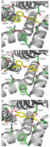
Comparison of the ligand binding pockets of the β1 and β2 adrenergic receptors. The ligand binding pockets are shown as viewed from the extracellular surface with EL2 removed for clarity (same colour scheme as in Fig. 1). (a) β2AR with the antagonist carazolol bound (PDB code 2RH1); (b) β1AR with the antagonist cyanopindolol bound (PDB code 2VT4); (c) β1AR with the agonist isoprenaline bound.
Figure 4
Figure 4
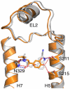
Differences in the ligand binding pocket between antagonist- and agonist-bound β1-adrenergic receptor. An alignment was performed (see Online Methods) between the structures of β1AR-m23 bound to either cyanopindolol (grey) or isoprenaline (orange) and the relative positions of the ligands and the transmembrane helices H5 and H7 are depicted. The 1 Å contraction of the ligand binding pocket between H5 and H7 is clear.

Comment in

References

    1. Evans BA, et al. Ligand-directed signalling at beta-adrenoceptors. Br J Pharmacol. 2010;159:1022–1038. - PMC - PubMed
    1. Rosenbaum DM, Rasmussen SG, Kobilka BK. The structure and function of G-protein-coupled receptors. Nature. 2009;459:356–363. - PMC - PubMed
    1. Ballesteros JA, Weinstein H. Integrated methods for the construction of three dimensional models and computational probing of structure function relations in G protein-coupled receptors. Methods Neurosci. 1995;25:366–428.
    1. Strader CD, et al. Conserved aspartic acid residues 79 and 113 of the beta-adrenergic receptor have different roles in receptor function. J Biol Chem. 1988;263:10267–10271. - PubMed
    1. Sato T, Kobayashi H, Nagao T, Kurose H. Ser203 as well as Ser204 and Ser207 in fifth transmembrane domain of the human beta2-adrenoceptor contributes to agonist binding and receptor activation. Br J Pharmacol. 1999;128:272–274. - PMC - PubMed

Publication types

MeSH terms