Review
. 2010 Dec 13;4(1):58-73.

Genetic and epigenetic alterations during renal carcinogenesis

Affiliations
Review

Genetic and epigenetic alterations during renal carcinogenesis

Eri Arai et al. Int J Clin Exp Pathol. .

Abstract

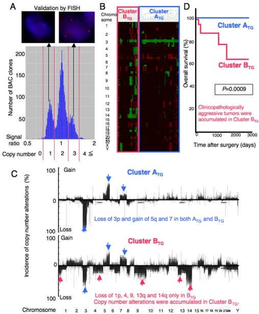
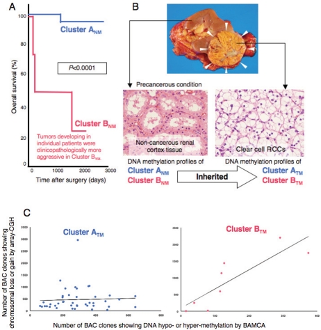
Renal cell carcinoma (RCC) is not a single entity, but comprises a group of tumors including clear cell RCC, papillary RCC and chromophobe RCC, which arise from the epithelium of renal tubules. The majority of clear cell RCCs, the major histological subtype, have genetic or epigenetic inactivation of the von Hippel-Lindau (VHL) gene. Germline mutations in the MET and fumarate hydratase (FH) genes lead to the development of type 1 and type 2 papillary RCCs, respectively, and such mutations of either the TSC1 or TSC2 gene increase the risk of RCC. Genome-wide copy number alteration analysis has suggested that loss of chromosome 3p and gain of chromosomes 5q and 7 may be copy number aberrations indispensable for the development of clear cell RCC. When chromosome 1p, 4, 9, 13q or 14q is also lost, more clinicopathologically aggressive clear cell RCC may develop. Since renal carcinogenesis is associated with neither chronic inflammation nor persistent viral infection, and hardly any histological change is evident in corresponding non-tumorous renal tissue from patients with renal tumors, precancerous conditions in the kidney have been rarely described. However, regional DNA hypermethylation on C-type CpG islands has already accumulated in such non-cancerous renal tissues, suggesting that, from the viewpoint of altered DNA methylation, the presence of precancerous conditions can be recognized even in the kidney. Genome-wide DNA methylation profiles in precancerous conditions are basically inherited by the corresponding clear cell RCCs developing in individual patients: DNA methylation alterations at the precancerous stage may further predispose renal tissue to epigenetic and genetic alterations, generate more malignant cancers, and even determine patient outcome. The list of tumor-related genes silenced by DNA hypermethylation has recently been increasing. Genetic and epigenetic profiling provides an optimal means of prognostication for patients with RCCs. Recently developed high-throughput technologies for genetic and epigenetic analyses will further accelerate the identification of key molecules for use in the prevention, diagnosis and therapy of RCCs.

Keywords: DNA methylation; Renal cell carcinoma; copy number alteration; precancerous condition; prognostication.


Figures

Figure 1
Figure 1
Macroscopic (A, C, F and G) and microscopic (B, D, E and H) views of a clear cell RCC (A and B), papillary RCCs (C, D and E) and a chromophobe RCC (F, G and H). A. Clear cell RCCs commonly protrude from the renal cortex as a rounded mass. Their cut surfaces are typically golden yellow, and necrosis and hemorrhage are commonly present. B. Clear cell RCCs typically have cytoplasm filled with lipids and glycogen and show an alveolar architecture. C. Papillary RCCs frequently contain areas of hemorrhage, necrosis and cystic degeneration. D. Type 1 papillary RCCs consist of papillae covered with a single or double layer of small cuboid cells with scanty cytoplasm. E. Type 2 papillary RCCs consist of papillae covered by large eosinophilic cells arranged in an irregular or pseudo-stratified manner. F. Chromophobe RCCs are solid circumscribed tumors with slightly lobulated surfaces. In unfixed specimens, the cut surface is homogeneously light brown or tan. G. Macroscopic view of the same chromophobe RCC after formalin fixation. The cut surface of chromophobe RCCs turns graysh-beige. H. Chromophobe RCCs consist of tumor cells with abundant eosinophilic cytoplasm (pale cells [Pa] and eosinophilic cells with a perinuclear halo [Eo]) and show mainly a solid structure.
Figure 2
Figure 2
Genetic clustering of clear cell renal cell carcinomas (RCCs). An example of a histogram of the signal ratios (test signal/reference signal) afforded by array-CGH in a clear cell RCC. The thresholds of the signal ratios for copy numbers of 0, 1, 2, 3 and 4 or more were determined from the troughs (red bars) between the distinct peaks. A. FISH analysis using the same clone validated the results of array-CGH (ref. 17). B. Unsupervised hierarchical clustering analysis based on array-CGH data. Clear cell RCCs were grouped into Clusters ATG and BTG (ref. 17). C. Distinct copy number profiles in Clusters ATG and BTG. LOSS of chromosome 3p and gain of 5q and 7 may promote the development of RCCs belonging to Cluster ATG and showing a favorable outcome. When loss of 1p, 4, 9, 13q or 14q is added, more malignant RCCs in Cluster BTG may develop (ref. 17). D. Kaplan-Meier survival curves based on genetic clustering of clear cell RCCs (Clusters ATG and BTG). None of the patients in Cluster ATG died as a result, and the overall survival rate of patients in Cluster BTG was significantly lower than that of patients in Cluster ATG (Log-rank test, ref. 17).
Figure 3
Figure 3
DNA methylation profiles in precancerous conditions and clear cell renal cell carcinomas (RCCs). A. Genome-wide DNA methylation profiles in the non-cancerous renal tissue were significantly correlated with clinicopathological parameters of clear cell RCCs developing in individual patients, and also outcome, indicating that DNA methylation alterations at the precancerous stage may generate more malignant cancers and even determine outcome (ref. 46). B. DNA methylation profiles in the non-cancerous renal tissue (Clusters ANM and BNM,see text) were basically inherited by the corresponding clear cell RCCs developing in individual patients as the DNA methylation profiles of Clusters ATM and BTM, respectively (ref. 46). C. In Cluster BTM, the number of clones showing copy number alterations by array-CGH was significantly correlated with that of DNA hypo- and hypermethylation by BAMCA in the same patient, whereas no such significant correlations were observed in Cluster ATM, suggesting that particular DNA methylation profiles may be closely related to chromosomal instability (unpublished data).

References

    1. Ferlay J, Shin HR, Bray F, Forman D, Mathers C, Parkin DM. Estimates of worldwide burden of cancer in 2008: GLOBOCAN 2008. Int J Cancer. in press; DOI: 10.1002/ijc.25516. - DOI - PubMed
    1. Pascual D, Borque A. Epidemiology of kidney cancer. Adv Urol. 2008 782381. - PMC - PubMed
    1. Baldewijns MM, van Vlodrop IJ, Schouten LJ, Soetekouw PM, de Bruine AP, van Engeland M. Genetics and epigenetics of renal cell cancer. Biochim Biophys Acta. 2008;1785:133–155. - PubMed
    1. Lopez-Beltran A, Carrasco JC, Cheng L, Scarpelli M, Kirkali Z, Montironi R. 2009 update on the classification of renal epithelial tumors in adults. Int J Urol. 2009;16:432–443. - PubMed
    1. Eble JN, Togashi K, Pisani P. Renal cell carcinoma. In “World Health Organization classification of tumours. Pathology and genetics. Tumours of the urinary system and male genital organs”. Lyon: IARC Press; 2004. pp. 10–43.

Publication types