. 2011 Apr;18(2):299-308.
doi: 10.1007/s12350-010-9331-2. Epub 2011 Jan 13.

Left-ventricular systolic and diastolic dyssynchrony as assessed by multi-harmonic phase analysis of gated SPECT myocardial perfusion imaging in patients with end-stage renal disease and normal LVEF

Affiliations

Left-ventricular systolic and diastolic dyssynchrony as assessed by multi-harmonic phase analysis of gated SPECT myocardial perfusion imaging in patients with end-stage renal disease and normal LVEF

Ji Chen et al. J Nucl Cardiol. 2011 Apr.

Abstract

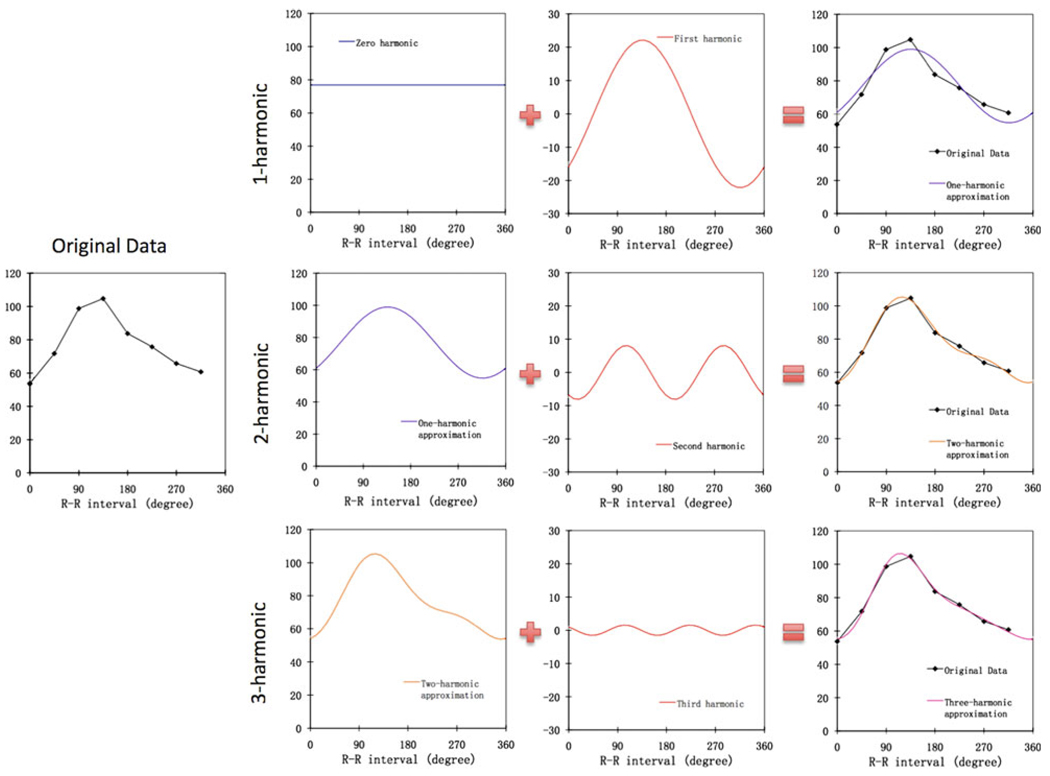
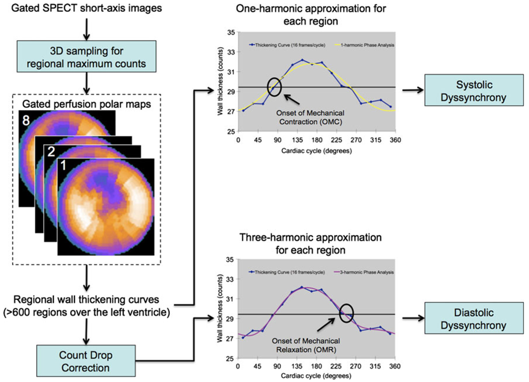
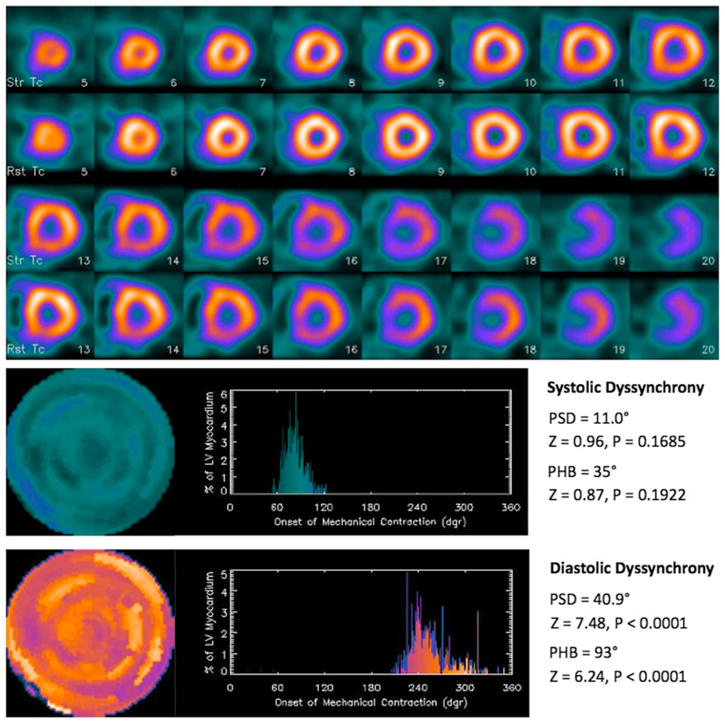
Background: The purpose of this study was to develop a multi-harmonic phase analysis method to measure diastolic dyssynchrony from conventional gated SPECT myocardial perfusion imaging (MPI) data and to compare it with systolic dyssynchrony in normal subjects and in patients with end-stage renal disease (ESRD) and normal left-ventricular ejection fraction (LVEF).

Methods: 121 consecutive patients with ESRD and normal LVEF and 30 consecutive normal controls were enrolled. Diastolic dyssynchrony parameters were calculated using 3-harmonic phase analysis. Systolic dyssynchrony parameters were calculated using the established 1-harmonic phase analysis.

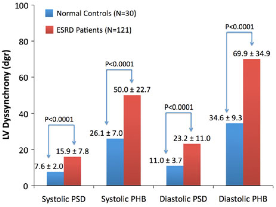
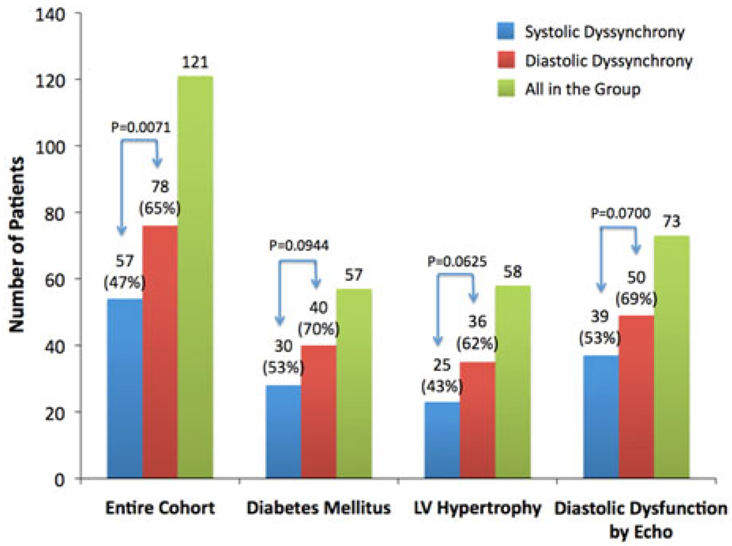
Results: The systolic and diastolic dyssynchrony parameters were correlated, but significantly different in both control and ESRD groups, indicating they were physiologically related but measured different LV mechanisms. The systolic and diastolic dyssynchrony parameters were each significantly different between the control and the ESRD groups. Significant systolic and diastolic dyssynchrony were found in 47% and 65% of the entire ESRD group.

Conclusion: Multi-harmonic phase analysis has been developed to assess diastolic dyssynchrony, which measured a new LV mechanism of regional function from gated SPECT MPI and showed a significantly higher prevalence rate than systolic dyssynchrony in patients with ESRD and normal LVEF.


Conflict of interest statement

The terms of this arrangement have been reviewed and approved by Emory University in accordance with its conflict-of-interest practice.

Figures

Figure 1
Figure 1
The use of the Fourier harmonics to approximate a typical discrete wall-thickening curve. The harmonic functions (zero-, first-, second-, and third-harmonic) were generated from the original data by the fast Fourier transform. The 1-harmonic approximation includes the zero- and first-harmonic functions. Adding the second- and third-harmonic functions to the approximation yields the 2- and 3-harmonic approximation, respectively. As shown in this figure, the lower harmonics determine the basic shape of the curve, whereas higher harmonics refine the approximation and improve its accuracy.
Figure 2
Figure 2
Processing steps of the multi-harmonic phase analysis tool. The input was the standard gated SPECT short-axis image. At first, regional maximal count detection was performed in 3D for each temporal frame to generate wall-thickening curves for over 600 LV regions. The wall-thickening curve for each region was approximated by the 1-harmonic function for systolic dyssynchrony and by the 3-harmonic function after a count drop correction for diastolic dyssynchrony. The count drop correction scaled every pixel in the last frame to make the total myocardial counts in the last frame equal to the total myocardial counts in the first frame. The phase angles derived by the systolic and diastolic approximation represented the onset of mechanical contraction (OMC) and onset of mechanical relaxation (OMR) respectively. Once the OMC and OMR phase angles of all regions were obtained, OMC and OMR phase distributions were generated that provided information on the degree of systolic and diastolic dyssynchrony for the entire LV, respectively.
Figure 3
Figure 3
Left-ventricular (LV) systolic and diastolic dyssynchrony parameters (PSD, phase standard deviation; PHB, phase histogram bandwidth) in normal controls vs patients with end-stage renal disease (ESRD) and normal LV ejection fraction (LVEF). All parameters were significantly different between the normal controls and ESRD patients by unpaired t test with unequal variance.
Figure 4
Figure 4
Prevalence rates of systolic and diastolic dyssynchrony in the entire ESRD group (N = 121) and the subsets of patients with diabetes mellitus (N = 57), and LV hypertrophy (N = 58) and LV diastolic dysfunction according to echocardiography (N = 73), respectively. Significant LV systolic and diastolic dyssynchrony were found in 47% and 65% the entire ESRD group, 53% and 70% of the patients with diabetes mellitus, 43% and 62% of the patients with echocardiographic evidence of LV hypertrophy, and 53% and 69% of the patients with echocardiographic evidence of diastolic dysfunction, respectively. There were no significant differences in prevalence rates of systolic and diastolic dyssynchrony between the entire ESRD group and each subset, indicating that these conditions did not result in higher prevalence of LV dyssynchrony in the ESRD patients than ESRD alone.
Figure 5
Figure 5
A patient example with significant diastolic dyssynchrony, but no significant systolic dyssynchrony. A 53-year-old male had end-stage renal disease, hypertension, diabetes mellitus, hemodialysis, but no coronary artery disease. He had LV hypertrophy and LVEF of 55% according to echocardiography. His QRS duration was 94 ms. Comparing his LV dyssynchrony parameters to the normal limits, he had no significant systolic dyssynchrony, but significant diastolic dyssynchrony by one-sample z test.

References

    1. Sarnak MJ, Levey AS, Schoolwerth AC, et al. Kidney disease as a risk factor for development of cardiovascular disease: A statement from the American Heart Association Councils on Kidney in Cardiovascular Disease, High Blood Pressure Research, Clinical Cardiology, and Epidemiology and Prevention. Circulation. 2003;108:2154–2169. - PubMed
    1. Verma A, Anavekar NS, Meris A, et al. The relationship between renal function and cardiac structure, function and prognosis after myocardial infarction. The VALIANT Echo Study. J Am Coll Cardiol. 2007;50:1238–1245. - PubMed
    1. Foley RN, Parfrey PS, Harnett JD, et al. Clinical and echocardiographic disease in patients starting end-stage renal disease therapy. Kidney Int. 1995;47:186–192. - PubMed
    1. Curtis BM, Parfrey PS. Congestive heart failure in chronic kidney disease: Disease-specific mechanisms of systolic and diastolic heart failure and management. Cardiol Clin. 2005;23:275–284. - PubMed
    1. Hayashi SY, Seeberger A, Lind B, Nowak J, do Nascimento MM, Lindholm B, et al. A single session of haemodialysis improves left ventricular synchronicity in patients with end-stage renal disease: A pilot tissue synchronization imaging study. Nephrol Dial Transplant. 2008;23:3622–3628. - PubMed

Publication types