

Microglial activation in regions related to cognitive function predicts disease onset in Huntington's disease: a multimodal imaging study
- PMID: 21229614
- PMCID: PMC6870088
- DOI: 10.1002/hbm.21008
Microglial activation in regions related to cognitive function predicts disease onset in Huntington's disease: a multimodal imaging study
Abstract
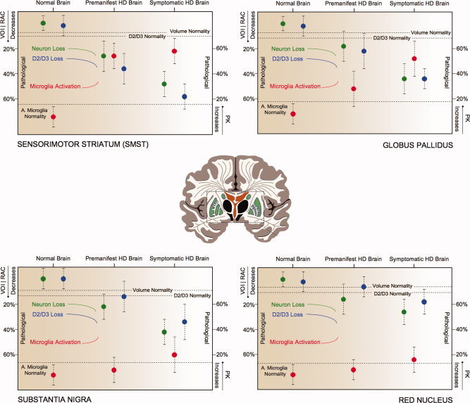
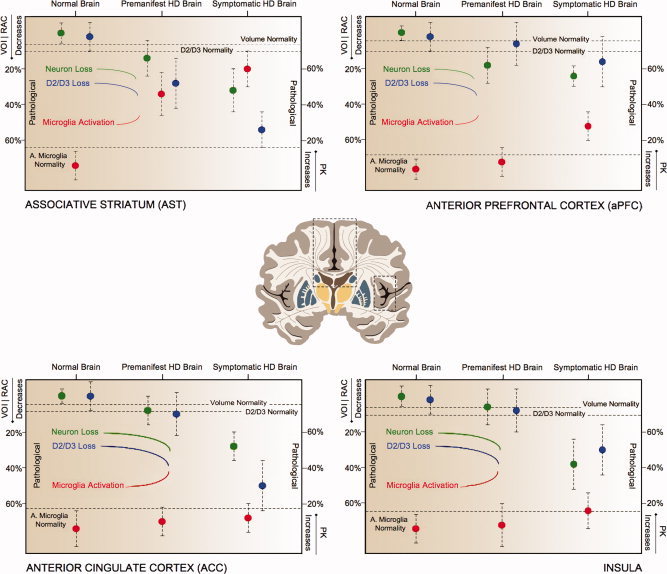
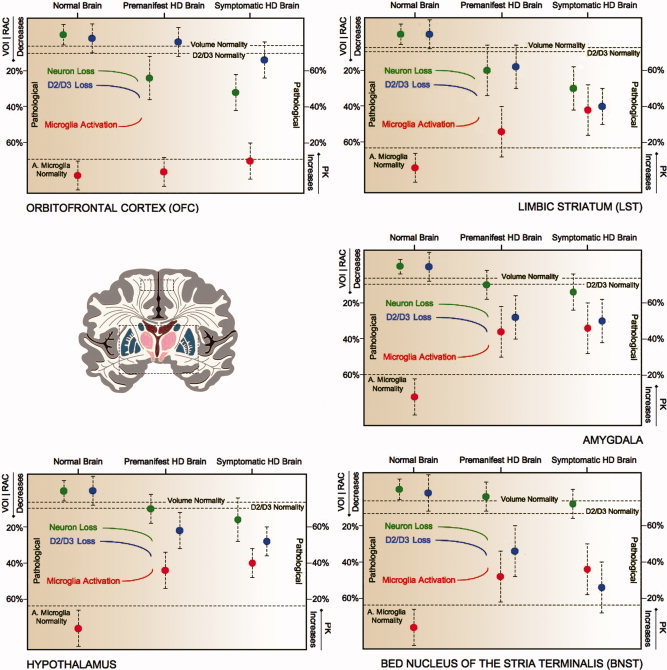
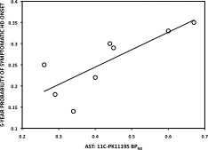
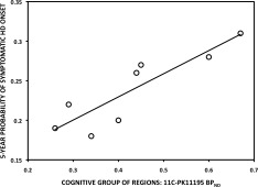
Huntington's disease (HD) is an inherited neurodegenerative disorder associated with motor, cognitive and psychiatric deficits. This study, using a multimodal imaging approach, aims to assess in vivo the functional and structural integrity of regions and regional networks linked with motor, cognitive and psychiatric function. Predicting disease onset in at risk individuals is problematic and thus we sought to investigate this by computing the 5-year probability of HD onset (p5 HD) and relating it to imaging parameters. Using MRI, (11)C-PK11195 and (11)C-raclopride PET, we have investigated volumes, levels of microglial activation and D2/D3 receptor binding in CAG repeat-matched groups of premanifest and symptomatic HD gene carriers. Findings were correlated with disease-burden and UHDRS scores. Atrophy was detected in sensorimotor striatum (SMST), substantia nigra, orbitofrontal and anterior prefrontal cortex in the premanifest HD. D2/D3 receptor binding was reduced and microglial activation increased in SMST and associative striatum (AST), bed nucleus of the stria terminalis, the amygdala and the hypothalamus. In symptomatic HD cases this extended to involve atrophy in globus pallidus, limbic striatum, the red nuclei, anterior cingulate cortex, and insula. D2/D3 receptor binding was additionally reduced in substantia nigra, globus pallidus, limbic striatum, anterior cingulate cortex and insula, and microglial activation increased in globus pallidus, limbic striatum and anterior prefrontal cortex. In premanifest HD, increased levels of microglial activation in the AST and in the regional network associated with cognitive function correlated with p5 HD onset. These data suggest that pathologically activated microglia in AST and other areas related to cognitive function, maybe better predictors of clinical onset and stresses the importance of early cognitive assessment in HD.
Copyright © 2010 Wiley-Liss, Inc.
Figures
References
-
- Andrews TC, Weeks RA, Turjanski N, Gunn RN, Watkins LH, Sahakian B, Hodges JR, Rosser AE, Wood NW, Brooks DJ ( 1999): Huntington's disease progression. PET and clinical observations. Brain 122: 2353–2363. - PubMed
-
- Banati RB ( 2002): Visualising microglial activation in vivo. GLIA 40: 206–217. - PubMed
-
- Björkqvist M, Wild EJ, Thiele J, Silvestroni A, Andre R, Lahiri N, Raibon E, Lee RV, Benn CL, Soulet D, Magnusson A, Woodman B, Landles C, Pouladi MA, Hayden MR, Khalili‐Shirazi A, Lowdell MW, Brundin P, Bates GP, Leavitt BR, Möller T, Tabrizi SJ ( 2008): A novel pathogenic pathway of immune activation detectable before clinical onset in Huntington's disease. J Exp Med 205: 1869–1877. - PMC - PubMed
-
- Bohanna I, Georgiou‐Karistianis N, Hannan AJ, Egan GF ( 2008): Magnetic resonance imaging as an approach towards identifying neuropathological biomarkers for Huntington's disease. Brain Res Rev 58: 209–225. - PubMed
-
- Bush G, Luu P, Posner MI ( 2000): Cognitive and emotional influences in anterior cingulate cortex. Trends Cogn Sci 4: 215–222. - PubMed
MeSH terms
Substances
Grants and funding
LinkOut - more resources
Full Text Sources
Other Literature Sources
Medical
Miscellaneous
