

Endothelial dysfunction of rat coronary arteries after exposure to low concentrations of mercury is dependent on reactive oxygen species
- PMID: 21232032
- PMCID: PMC3081124
- DOI: 10.1111/j.1476-5381.2011.01203.x
Endothelial dysfunction of rat coronary arteries after exposure to low concentrations of mercury is dependent on reactive oxygen species
Abstract
Background and purpose: Exposure to mercury is known to increase cardiovascular risk but the underlying mechanisms are not well explored. We analysed whether chronic exposure to low mercury doses affects endothelial modulation of the coronary circulation.
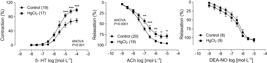
Experimental approach: Left coronary arteries and hearts from Wistar rats treated with either HgCl(2) (first dose 4.6 µg·kg(-1) , subsequent doses 0.07 µg·kg(-1) day(-1) , 30 days) or vehicle were used. Endothelial cells from pig coronary arteries incubated with HgCl(2) were also used.
Key results: Mercury treatment increased 5-HT-induced vasoconstriction but reduced acetylcholine-induced vasodilatation. It also reduced nitric oxide (NO) production and the effects of NO synthase inhibition with L-NAME (100 µmol·L(-1) ) on 5-HT and acetylcholine responses. Superoxide anion production and mRNA levels of NOX-1 and NOX-4 were all increased. The superoxide anion scavenger tiron (1 mmol·L(-1)) reduced 5-HT responses and increased acetylcholine responses only in vessels from mercury-treated rats. In isolated hearts from mercury-treated rats, coronary perfusion and diastolic pressure were unchanged, but developed isovolumetric systolic pressure was reduced. In these hearts, L-NAME increased coronary perfusion pressure and diastolic pressure while it further reduced developed systolic pressure.
Conclusions and implications: Chronic exposure to low doses of mercury promotes endothelial dysfunction of coronary arteries, as shown by decreased NO bioavailability induced by increased oxidative stress. These effects on coronary function increase resistance to flow, which under overload conditions might cause ventricular contraction and relaxation impairment. These findings provide further evidence that mercury, even at low doses, could be an environmental risk factor for cardiovascular disease.
© 2011 The Authors. British Journal of Pharmacology © 2011 The British Pharmacological Society.
Figures


Similar articles
-
Apocynin prevents vascular effects caused by chronic exposure to low concentrations of mercury.PLoS One. 2013;8(2):e55806. doi: 10.1371/journal.pone.0055806. Epub 2013 Feb 4. PLoS One. 2013. PMID: 23390552 Free PMC article.
-
Low mercury concentrations cause oxidative stress and endothelial dysfunction in conductance and resistance arteries.Am J Physiol Heart Circ Physiol. 2008 Sep;295(3):H1033-H1043. doi: 10.1152/ajpheart.00430.2008. Epub 2008 Jul 3. Am J Physiol Heart Circ Physiol. 2008. PMID: 18599595
-
Low mercury concentration produces vasoconstriction, decreases nitric oxide bioavailability and increases oxidative stress in rat conductance artery.PLoS One. 2012;7(11):e49005. doi: 10.1371/journal.pone.0049005. Epub 2012 Nov 7. PLoS One. 2012. PMID: 23145049 Free PMC article.
-
Cerebrovascular endothelial dysfunction induced by mercury exposure at low concentrations.Neurotoxicology. 2016 Mar;53:282-289. doi: 10.1016/j.neuro.2016.02.010. Epub 2016 Mar 2. Neurotoxicology. 2016. PMID: 26945730
-
Persistence of EDHF pathway and impairment of the nitric oxide pathway after chronic mercury chloride exposure in rats: mechanisms of endothelial dysfunction.Hum Exp Toxicol. 2011 Nov;30(11):1777-84. doi: 10.1177/0960327110391389. Epub 2010 Dec 9. Hum Exp Toxicol. 2011. PMID: 21148200
Cited by
-
Toxic effects of mercury on the cardiovascular and central nervous systems.J Biomed Biotechnol. 2012;2012:949048. doi: 10.1155/2012/949048. Epub 2012 Jul 2. J Biomed Biotechnol. 2012. PMID: 22811600 Free PMC article. Review.
-
Ferroptosis as a mechanism of non-ferrous metal toxicity.Arch Toxicol. 2022 Sep;96(9):2391-2417. doi: 10.1007/s00204-022-03317-y. Epub 2022 Jun 21. Arch Toxicol. 2022. PMID: 35727353 Review.
-
Transition metals in angiogenesis - A narrative review.Mater Today Bio. 2023 Aug 3;22:100757. doi: 10.1016/j.mtbio.2023.100757. eCollection 2023 Oct. Mater Today Bio. 2023. PMID: 37593220 Free PMC article. Review.
-
Apocynin prevents vascular effects caused by chronic exposure to low concentrations of mercury.PLoS One. 2013;8(2):e55806. doi: 10.1371/journal.pone.0055806. Epub 2013 Feb 4. PLoS One. 2013. PMID: 23390552 Free PMC article.
-
Re-thinking the link between exposure to mercury and blood pressure.Arch Toxicol. 2025 Feb;99(2):481-512. doi: 10.1007/s00204-024-03919-8. Epub 2025 Jan 3. Arch Toxicol. 2025. PMID: 39804370 Free PMC article.
References
-
- ATSDR. Agency for Toxic Substances and Disease Registry. National Alert. A warning about continuing patterns of metallic mercury exposure. 1997. Available at: http://www.atsdr.cdc.gov/alerts/970626.html.
-
- Björkman L, Sandborgh-Englund G, Ekstrand J. Mercury in saliva and feces after removal of amalgam fillings. Toxicolol Appl Pharmacol. 1997;144:156–162. - PubMed
-
- Chen C, Qu L, Li B, Xing L, Jia G, Wang T, et al. Increased oxidative DNA damage, as assessed by urinary 8-hydroxy-2′-deoxyguanosine concentrations, and serum redox status in persons exposed to mercury. Clin Chem. 2005;51:759–767. - PubMed
-
- Drexler H, Hornig B. Endothelial dysfunction in human disease. J Mol Cell Cardiol. 1999;31:51–60. - PubMed
Publication types
MeSH terms
Substances
LinkOut - more resources
Full Text Sources
