. 2011 Jan 13;8(1):3.
doi: 10.1186/1742-2094-8-3.

Microglial MAC1 receptor and PI3K are essential in mediating β-amyloid peptide-induced microglial activation and subsequent neurotoxicity

Affiliations

Microglial MAC1 receptor and PI3K are essential in mediating β-amyloid peptide-induced microglial activation and subsequent neurotoxicity

Dan Zhang et al. J Neuroinflammation. .

Abstract

Background: β-Amyloid peptide (Aβ) is a major protein in the brain associated with Alzheimer's and Parkinson's diseases. The purpose of this study was to investigate the role of macrophage antigen-1 (MAC1) receptor, an integrin scavenger receptor in microglia, and subsequent signaling events in mediating Aβ-induced neurotoxicity. We have previously reported that NADPH oxidase (PHOX) on microglia and superoxide produced by PHOX are critical for Aβ-induced loss of dopaminergic neurons. However, the upstream signaling pathway of superoxide production remains unclear.

Methods: For the in vitro study, mesencephalic neuron-glia cultures and microglia-enriched cultures from mice deficient in the MAC1 receptor (MAC1-/-) and wild type controls were used to investigate the role of MAC1 receptor in Aβ-induced neurotoxicity and the role of phosphoinositide-3 kinase (PI3K) in the signal pathway between MAC1 receptor and PHOX. For the in vivo study, Aβ was injected into the substantia nigra of MAC1-/- mice and wild type mice to confirm the role of MAC1 receptor.

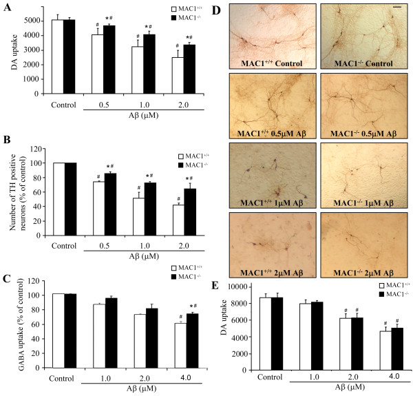
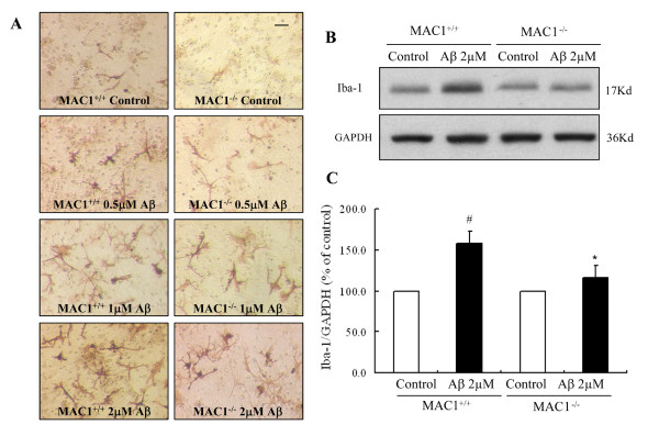
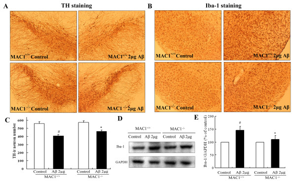
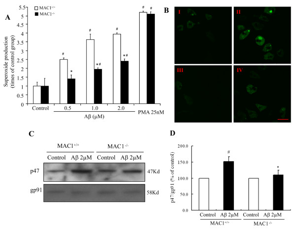
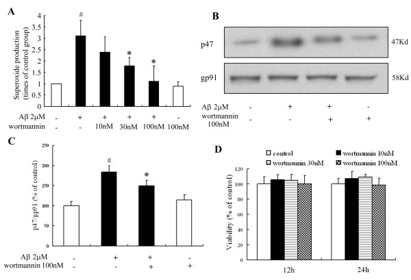
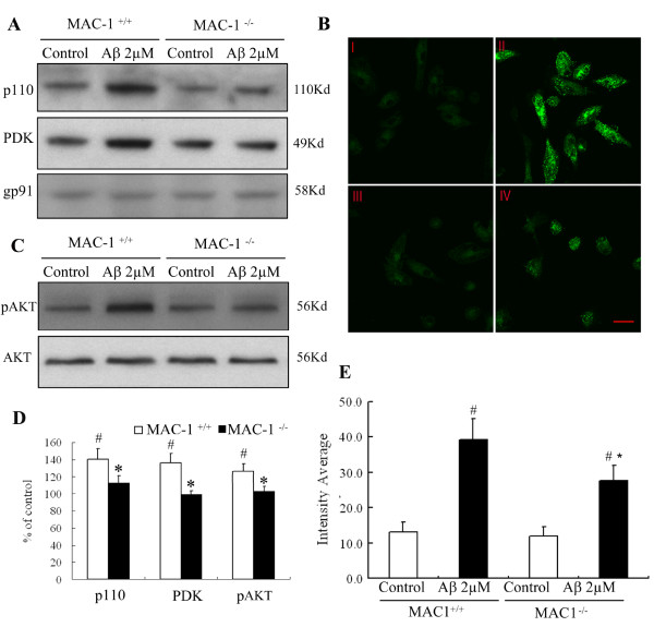
Results: We found that Aβ-induced activation of microglia, activation of PHOX, generation of superoxide and other reactive oxygen species, and loss of dopaminergic neurons were decreased in MAC1-/- cultures compared to MAC1+/+ cultures. In MAC1-/- mice, dopaminergic neuron loss in response to Aβ injection into the substantia nigra was reduced relative to MAC1+/+ mice. Thus, MAC1 receptor-mediated PHOX activation and increased superoxide production are associated with Aβ-induced neurotoxicity. PI3K activation was one downstream step in MAC1 signaling to PHOX and played an important role in Aβ-induced neurotoxicity. In microglia-enriched cultures from MAC1-/- mice, Aβ-induced activation of PI3K (phosphorylation of target proteins and PIP3 production) was reduced relative to MAC1+/+ cultures.

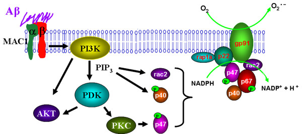
Conclusions: Taken together, our data demonstrate that Aβ activates MAC1 receptor to increase the activity of PI3K, which in turn phosphorylates p47phox, triggers the translocation of cytosolic subunits of PHOX to microglia membrane, increases PHOX activation and the subsequent production of superoxide and causes neurotoxicity.


Figures

Figure 1
Figure 1
Absence of MAC1 attenuates Aβ-induced dopaminergic and GABAergic neurotoxicity in neuron-glia cultures. Mice (MAC1+/+ and MAC1-/-) mesencephalic neuron-glia cultures in 24-well plates were treated with vehicle medium (control group) or 0.5 μM, 1.0 μM and 2.0 μM Aβ for 7 days. (A) Aβ-induced dopaminergic neurotoxicity was quantified by [3H] dopamine uptake assay. (B) Numbers of TH-positive cells remaining in the neuron-glia cultures after vehicle or Aβ treatment. (D) Representative light microscopic images are shown for TH-positive neurons treated with vehicle or Aβ. Scale bar: 100 μm. (C) Mice (MAC1+/+ and MAC1-/-) cortical neuron-glia cultures in 24-well plates were treated with vehicle or 1.0 μM, 2.0 μM and 4.0 μM Aβ for 7 days. Aβ-induced GABAergic neurotoxicity was quantified by [3H] GABA uptake assay. (E) Mice (MAC1+/+ and MAC1-/-) mesencephalic microglia-depleted cultures in 24-well plates were treated with vehicle medium (control group) or 1.0 μM, 2.0 μM and 4.0 μM Aβ for 7 days. Aβ-induced dopaminergic neurotoxicity was quantified by [3H] dopamine uptake assay. Results are from four independent experiments. #: p < 0.05 compared with corresponding vehicle-treated controls. *: p < 0.05 compared with MAC1+/+ cultures after same treatments.
Figure 2
Figure 2
MAC1 is necessary for Aβ-induced microglial activation. Mesencephalic neuron-glia cultures from MAC1+/+ and MAC1-/- mice were treated with vehicle medium (control group) or Aβ for 2 days. (A) Cultures were fixed after treatments. Microglia were visualized by immunostaining of the F4/80 antigen, a microglial marker. The images presented are representative of three independent experiments. Scale bar: 50 μm. (B) Western blot analysis of microglial activation. Cell lysates of cultures from MAC1+/+ and MAC1-/- mice were prepared 2 days after vehicle or 2.0 μM Aβ treatment and immunoblot analysis was performed for the measurement of Iba1 antigen. GAPDH was used as loading control. (C) The ratio of densitometry values of Iba1 and GAPDH was analyzed and normalized to respective control. Results are presented as mean ± SEM for three independent experiments. #: p < 0.05 compared with corresponding vehicle-treated controls. *: p < 0.05 compared with MAC1+/+ cultures after same treatments.
Figure 3
Figure 3
Dopaminergic neurons from MAC1-deficient mice are more resistant to Aβ-induced neurotoxicity in vivo. Two μg of Aβ were injected into the right side of adult MAC1+/+ and MAC1-/- mouse SN; vehicle saline alone (control group) was injected into the left side. After 7 days, brains were removed, dopaminergic neurons were stained with an antibody against TH and the microglia were stained with an antibody against Iba-1. Ten animals were used for each group. Dopaminergic neurons were counted in a double-blind manner by three individuals. (A) Immunocytochemical analysis of TH-positive neurons. (B) Number of TH-positive neurons. (C) Immunocytochemical analysis of microglia. (D) Western blot analysis of microglial activation in midbrain area. GAPDH was used as loading control. (E) The ratio of densitometry values of Iba1 and GAPDH was analyzed and normalized to respective control. Results are presented as mean ± SEM. #: p < 0.05 compared with corresponding saline-treated side. *: p < 0.05 compared with MAC1+/+ mice after same treatments.
Figure 4
Figure 4
MAC1 mediates activation of PHOX and production of superoxide. (A) Microglia-enriched cultures from MAC1+/+ and MAC1-/- mice were treated with vehicle medium (control group), Aβ or PMA for 10 min. Extracellular superoxide generation was measured by the SOD-inhibitable reduction of tetrazolium salt, WST-1. (B) Microglia-enriched cultures from MAC1+/+ and MAC1-/- mice were incubated with HBSS containing 5 μM DCFDA in the dark for 1 hour. Then cells were treated with vehicle medium (control group) or Aβ for 10 min. Fluorescent images were captured using Zeiss 510 laser scanning confocal microscope. Scale bar: 25 μm. (C) Western blot assays for p47phox levels in membrane fractions of microglia from MAC1+/+ and MAC1-/- mice 10 min after vehicle medium (control group) or Aβ treatment. (D) Densitometry analysis was performed with values of p47phox normalized to loading control and further normalized to control levels. Data are presented as mean ± SEM from three independent experiments. #: p < 0.05 relative to corresponding vehicle-treated control cultures. *: p < 0.05 relative to MAC1+/+ cultures after same treatments.
Figure 5
Figure 5
PI3K mediates signaling between MAC1 receptor and PHOX. Microglia-enriched cultures from MAC1+/+ mice were pretreated with wortmannin for 30 min and then challenged with Aβ for 10 min. (A) Extracellular superoxide generation was measured by the SOD-inhibitable reduction of tetrazolium salt, WST-1. (B) Western blot assays for p47phox levels in membrane fractions from microglia. (C) Densitometry analysis was performed with values of p47phox normalized to loading control and further normalized to control levels. (D) Microglia-enriched cultures from MAC1+/+ mice were treated with vehicle medium (control group) or different concentrations of wortmannin for 12 or 24 hours; cell viability was measured using MTT assay. Data are presented as mean ± SEM for three independent experiments. #: p < 0.05 relative to vehicle-treated control cultures. *: p < 0.05 relative to Aβ-treated cultures.
Figure 6
Figure 6
MAC1 is necessary for PI3K activation elicited by Aβ. (A) Western blot assays for p110 and PDK levels in membrane fractions of microglia from MAC1+/+ and MAC1-/- mice 10 min after vehicle medium (control group) or Aβ treatment. (B) Enriched microglial cells from MAC1+/+ and MAC1-/- mice were treated with vehicle medium (control group) or Aβ for 10 min, fixed and permeabilized. I and II are MAC1+/+ cultures treated with vehicle or Aβ, respectively. III and IV are MAC1-/- cultures treated with vehicle or Aβ, respectively. (C) Western blot assays for pAKT and AKT levels in microglia from MAC1+/+ and MAC1-/- mice 10 min after vehicle medium (control group) or Aβ treatment. (D) Densitometry was performed with values normalized to respectively loading control and further normalized to control levels. Cells were incubated with a rabbit polyclonal antibody against PIP3 and then with a FITC-conjugated goat anti-rabbit antibody. Shown are representative confocal images. Scale bar: 25 μm. (E) Quantitative analysis of the figures in 6B. Average cellular fluorescence was quantitated from at least 100 separate cellular measurements obtained from each treatment group. Background fluorescence, determined in the absence of fluorescence labeled antibody, was minimal in both cell populations (data not shown). Experiments were performed at least three times. #: p < 0.05 relative to corresponding vehicle-treated control cultures. *: p < 0.05 relative to MAC1+/+ cultures after same treatments.
Figure 7
Figure 7
β-amyloid signals through microglial MAC1 receptor and PI3K to PHOX. Microglia exposed to Aβ exhibit a respiratory burst leading to the release of superoxide anion (O2-). Release of superoxide is mediated in part through the Aβ cell surface receptor MAC1. Aβ engagement of MAC1 receptor leads to the initiation of complex signaling events mediated by PI3K activation and PIP3 production, which leads to AKT phosphorylation and activation of PDK. Activation of these signaling cascades is linked to the activation of PHOX. PHOX plays an essential role in innate immunity by catalyzing the formation of superoxide. PHOX consists of two integral membrane proteins, p22phox and gp91phox, which together form a heterodimeric flavoprotein known as cytochrome b558. In addition, there are four cytosolic components p47phox, p67phox, p40phox, and the small G-protein Rac. As an important component for PHOX activation, the GDP/GTP exchange on Rac-1 is reported to be a point of possible PI3K intervention. PIP3 is reported to bind to p47phox and p40phox, and mediates their phosphorylation[46]; PI3K signaling pathway may also sometimes be involved in PKC activation, thus play an important role in the phosphorylation of p47phox. The cytosolic components then translocate to the membrane where they form a complex with cytochrome b558. The oxidase complex then initiates electron flow and generation of superoxide through the NADPH-derived electron reduction by the flavocytochrome. These findings suggest that MAC1 and PI3K are involved in upstream signaling cascades responsible for activating PHOX assembly and microglia in response to Aβ.

References

    1. Arai H, Lee VM, Hill WD, Greenberg BD, Trojanowski JQ. Lewy bodies contain beta-amyloid precursor proteins of Alzheimer's disease. Brain Res. 1992;585:386–390. doi: 10.1016/0006-8993(92)91242-7. - DOI - PubMed
    1. Yoshida T, Ohno-Matsui K, Ichinose S, Sato T, Iwata N, Saido TC, Hisatomi T, Mochizuki M, Morita I. The potential role of amyloid beta in the pathogenesis of age-related macular degeneration. J Clin Invest. 2005;115:2793–2800. doi: 10.1172/JCI24635. - DOI - PMC - PubMed
    1. Jendroska K, Kashiwagi M, Sassoon J, Daniel SE. Amyloid beta-peptide and its relationship with dementia in Lewy body disease. J Neural Transm Suppl. 1997;51:137–144. - PubMed
    1. Haga S, Akai K, Ishii T. Demonstration of microglial cells in and around senile (neuritic) plaques in the Alzheimer brain. An immunohistochemical study using a novel monoclonal antibody. Acta Neuropathol. 1989;77:569–575. doi: 10.1007/BF00687883. - DOI - PubMed
    1. Itagaki S, McGeer PL, Akiyama H, Zhu S, Selkoe D. Relationship of microglia and astrocytes to amyloid deposits of Alzheimer disease. J Neuroimmunol. 1989;24:173–182. doi: 10.1016/0165-5728(89)90115-X. - DOI - PubMed

Publication types

MeSH terms

LinkOut - more resources