. 2011 Jan 13;4(1):3.
doi: 10.1186/1755-1536-4-3.

Hyaluronidase recruits mesenchymal-like cells to the lung and ameliorates fibrosis

Affiliations

Hyaluronidase recruits mesenchymal-like cells to the lung and ameliorates fibrosis

Claudia S Bitencourt et al. Fibrogenesis Tissue Repair. .

Abstract

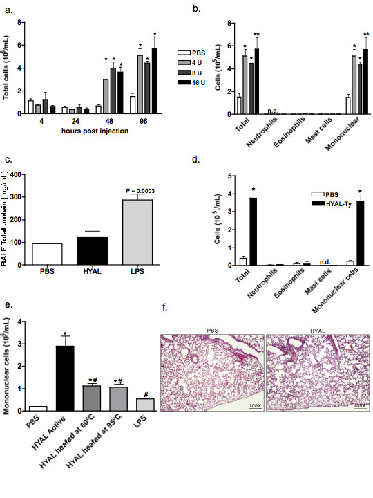
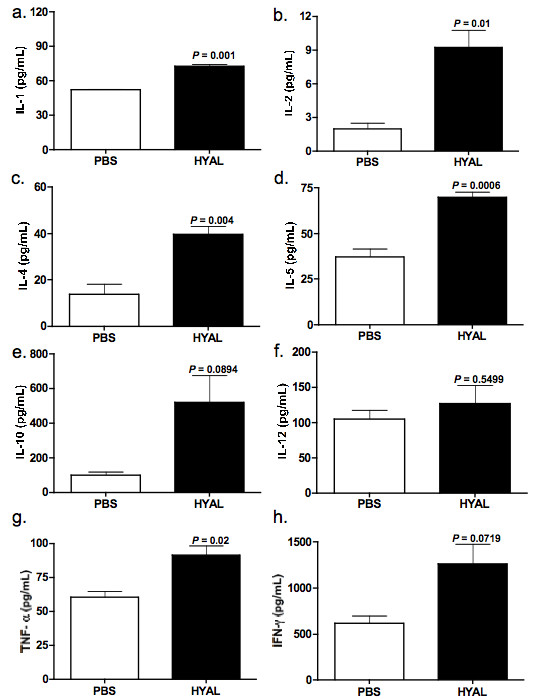
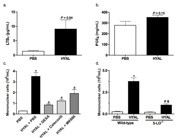
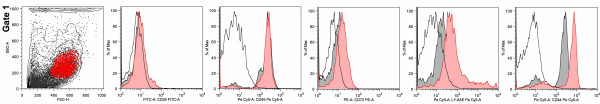
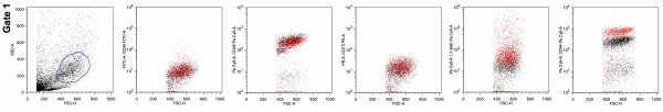
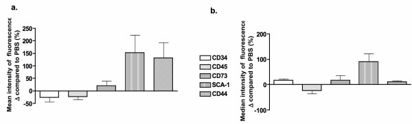
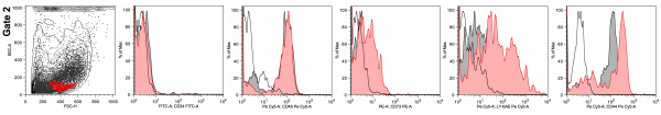
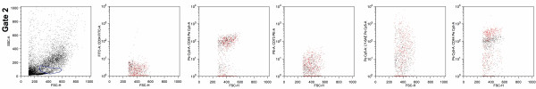
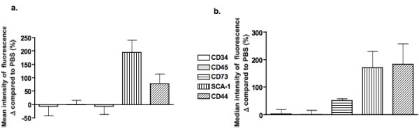
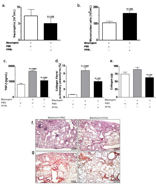
Hyaluronidases (HYALs) comprise a group of enzymes that degrade hyaluronic acid (HA). In this report, we reveal that a single intranasal inoculation of HYAL induces an increase in mononuclear cells within the bronchoalveolar space demonstrating a mesenchymal-like phenotype, expressing stem cell antigen-1 (SCA-1), CD44 and CD73 but not CD34, CD45, CD3, CD4, CD8 or CD19. This influx of mesenchymal stem cell (MSC)-like cells was dependent on leukotriene production within the lung parenchyma. These findings prompted experiments demonstrating that HYAL treatment potently blocked bleomycin-induced lung injury and fibrosis while decreasing transforming growth factor (TGF)-β production and collagen deposition. These data suggest that HYAL is a novel and promising tool to use autologous MSC-like cells in the treatment of pulmonary fibrosis.


Figures

Figure 1
Figure 1
Hyaluronidase (HYAL) induces an increase in leukocytes in bronchoalveolar fluid (BALF) of C57Bl/6 mice. (A) Total cells obtained from bronchoalveolar space after 4, 24, 48 and 96 h of inoculation with 4, 8 or 16 U of bovine testicular HYAL. Values represent means ± standard error of mean (SEM); n = 4; * P < 0.05 compared to phosphate buffered saline (PBS); ANOVA test was used. (B) HYAL induces almost exclusively mononuclear cell increases in the bronchoalveolar space after 96 h of inoculation with 16 U of bovine testicular HYAL in C57Bl/6 mice. N.d = not detected. Values represent means ± SEM; n = 4; * P < 0.05, ** P < 0.01 compared to PBS by ANOVA. (C) 16 U HYAL did not induce oedema after 96 h. The total protein content was measured in BALF. Llopopolysaccharide was inoculated intranasally (i.n.) as a positive control of oedema (500 μg/μL in 20 μL). Values represent means ± SEM; n = 4; P = 0.003 t test compared to the PBS. (D) Tityus serrulatus hyaluronidase (HYAL-TS) also induced mononuclear increase in the bronchoalveolar space after i.n. inoculation of 16 U in C57Bl/6 mice. N.d = not detected. Values represent means ± SEM; n = 5; * P = 0.003 using test t, compared to the PBS. (E) HYAL inactivation by heating reduces mononuclear cell numbers in bronchoalveolar space. 16 U of bovine testicular HYAL were heated or not at 60°C or 95°C i.n. inoculated and BALF cells collected 96 h later. Values represent means ± SEM; n = 5; * P < 0.001 compared to PBS; # P < 0.001 compared to active HYAL by ANOVA. (F) Photomicrographs of representative lung sections obtained from mice inoculated with PBS or HYAL. Tissues were stained with H&E to investigate inflammatory cells accumulation. Original magnification: 100x.
Figure 2
Figure 2
Hyaluronidase (HYAL) modulates cytokines in the lung parenchyma. Supernatants were prepared from the lung parenchyma obtained from C57Bl/6 mice treated after 96 h of inoculation with 16 U of bovine testicular HYAL or phosphate buffered saline (PBS). Interleukin (IL)-1, IL-2, IL-4, IL-5, IL-10, IL-12, tumour necrosis factor (TNF)-α and interferon (IFN)-γ were assayed by ELISA. (A) IL-1, P = 0.001; (B) IL-2, P = 0.01; (C) IL-4, P = 0.004; (D) IL-5, P = 0.006; (E) IL-10, P = 0.08; (F) IL-12, P = 0.54; (G) TNF-α, P = 0.02; (H) IFN-γ, P = 0.07; (A-H) = values represent means ± SEM compared to PBS; n = 5, t test was used.
Figure 3
Figure 3
Hyaluronidase (HYAL) induces mononuclear cell increase in bronchoalveolar space by mechanism partially dependent on lipid mediators. (A) Leukotriene (LT)B4 presents in the supernatant of lung homogenates were analysed by specific electroimmunoassay. C57Bl/6 was inoculated intranasally (i.n.) with 16 U of bovine testicular HYAL or phosphate buffered saline (PBS) and the lungs were removed 96 h later. Values represent means ± standard error of mean (SEM); n = 10; P = 0.04 compared to the PBS; t test was used. (B) Prostaglandin E2 concentration in the supernatant of lung homogenates was analysed by specific electroimmunoassay. C57Bl/6 was inoculated i.n. with 16 U of bovine testicular HYAL or PBS and the lung was removed 96 h later. Values represent means ± SEM; n = 10; P = 0.15 compared to the PBS group, t test was used. (C) Pre-treatment with dexamethasone (1 mg/kg s.c.); MK886 (5 mg/kg/0.5 mL by gavage) or celecoxib (5 mg/kg/0.5 mL by gavage) significantly reduced the number of cells in bronchoalveolar space of C57Bl/6 mice. All treatments were performed 1 h before stimuli and every 24 h until the end of experiment. Control received only PBS. Values represent means ± SEM.; n = 5; * P > 0.01 compared to PBS; # P > 0.001 compared to HYAL + PBS, ANOVA test was used. (C) Mononuclear cells recruitment to the bronchoalveolar space of 5-LO -/- (129-Alox5tm1Fun) mice is decrease when compared to strain-matched wild-type (WT) mice. Each strain was inoculated with 16 U of HYAL and cells recovered 96 h after stimulus. Values represent means ± SEM; n = 5; * P < 0.0001 compared to PBS WT; # P = 0.0003 compared to PBS 5-LO -/-; # P = 0.0003 compared to HYAL WT; test t was used.
Figure 4
Figure 4
Hyaluronidase (HYAL) induced an increase in bronchoalveolar cells with a phenotype of mesenchymal stem cells. Gate 1 is a representative picture showing forward/side scatters dot-plot of bronchoalveolar fluid cells obtained from phosphate buffered saline (PBS) treatment (black contour-plot) overlaid upon HYAL exposure (grey dot-plot), as well as the gate population highlighted in red. Histograms overlays were done using the gated population highlighted in red; open histogram = isotype; grey filled histogram = PBS; red filled histogram = HYAL. The figure depicts a representative analysis from five independent experiments.
Figure 5
Figure 5
Hyaluronidase (HYAL) induced an increase in bronchoalveolar cells with a phenotype of mesenchymal stem cells. Gate 1 is a representative picture showing forward/side scatters dot-plot of bronchoalveolar cells obtained from PBS treatment (black) overlaid upon HYAL exposure (red). The figure depicts a representative analysis from five independent experiments.
Figure 6
Figure 6
Hyaluronidase (HYAL) induced an increase in bronchoalveolar cells with a phenotype of mesenchymal stem cells. The mean (A) and median (B) expression of markers is altered by HYAL in gate 1. Phosphate buffered saline values were considered to be 100%. The figure depicts a representative analysis from five independent experiments.
Figure 7
Figure 7
Hyaluronidase (HYAL) induced an increase in bronchoalveolar cells with a phenotype of mesenchymal stem cells. Gate 2 is a representative picture showing forward/side scatters dot-plot of bronchoalveolar fluid cells obtained from phosphate buffered saline (PBS) treatment (black contour-plot) overlaid upon HYAL exposure (grey dot-plot), as well as the gate highlighted in red. Histograms overlays were done using the gated population highlighted in red; open histogram = isotype; grey filled histogram = PBS; red filled histogram = HYAL. The figure depicts a representative analysis from five independent experiments.
Figure 8
Figure 8
Hyaluronidase (HYAL) induced an increase in bronchoalveolar cells with a phenotype of mesenchymal stem cells. Gate 2 is a representative picture showing forward/side scatters dot-plot of bronchoalveolar fluid cells obtained from phosphate buffered saline treatment (black) overlaid upon HYAL exposure (red). Figure depicts a representative analysis from five independent experiments.
Figure 9
Figure 9
Hyaluronidase (HYAL) induced an increase in bronchoalveolar cells with a phenotype of mesenchymal stem cells. The mean (A) and median (B) expression of markers was altered by HYAL in gate 2, presented as the percentage of variation compared to the control. The controls mice were considered to be 100%. The figure depicts a representative analysis from five independent experiments.
Figure 10
Figure 10
Effects of hyaluronidase (HYAL) treatment on bleomycin-induced inflammation in the lung. At day 8 after bleomycin treatment (4 U/kg/0.1 mL NaCl 0.9%) in the lungs of C57Bl/6 mice, animals were treated with intranasal inoculation (i.n.) once with 16 U HYAL or phosphate buffered saline (PBS) and inflammatory cells recovered 96 h after. (A) Neutrophils numbers in the bleomycin-inoculated lung were not altered by HYAL (P = 0.406). However, (B) mononuclear cells were significantly increased by HYAL treatment (P = 0.045). Values represent means ± standard error of mean (SEM).; n = 5, by t test (C) transforming growth factor (TGF)-β is increased by bleomycin but reduced by HYAL treatment. TGF-β was measured in supernatant of lung tissue homogenate by ELISA. Values represent means ± SEM; n = 5, P = 0.0024 compared to bleomycin, P < 0.0001 compared to PBS, by t test. (D) The picrosirius morphometric analysis corresponding to the area occupied by the fibres were determined by digital densitometry recognition and expressed as a percentage of the total area of the field. Values represent means ± SEM; n = 10; P = 0.010 compared to PBS treated, using t test. (E) Acid-soluble collagen in the supernatant of lung tissue homogenate, assayed by Sircol. Values represent means ± SEM; n = 10; P = 0.010 compared to PBS, by t test. (F, G) Photomicrographs of representative lung sections obtained from mice inoculated with bleomycin and treated with PBS or with HYAL. In (F) tissues were stained with haematoxylin and eosin in order to investigate inflammatory cells accumulation and in (G) with Picrosirius to determine the collagen content. The histological analyses were single-blinded. Original magnifications = 100x.

Similar articles

Cited by

References

    1. Kemparaju K, Girish KS. Snake venom hyaluronidase: a therapeutic target. Cell Biochem Funct. 2006;24:7–12. doi: 10.1002/cbf.1261. - DOI - PubMed
    1. Hynes WL, Dixon AR, Walton SL, Aridgides LJ. The extracellular hyaluronidase gene (hylA) of streptococcus pyogenes. FEMS Microbiol Lett. 2000;184:109–112. doi: 10.1111/j.1574-6968.2000.tb08999.x. - DOI - PubMed
    1. Jiang D, Liang J, Noble PW. Hyaluronan in tissue injury and repair. Annu Rev Cell Dev Biol. 2007;23:435–461. doi: 10.1146/annurev.cellbio.23.090506.123337. - DOI - PubMed
    1. Menzel EJ, Far C. Hyaluronidase and its substrate: biochemistry, biological activities and therapeutic uses. Cancer Lett. 1998;131:3–11. doi: 10.1016/S0304-3835(98)00195-5. - DOI - PubMed
    1. Savani RC, Hou G, Liu P, Wang C, Simons E, Grimm PC, Stern R, Greenberg AH, DeLisser HM, Khalil N. A role for hyaluronan in macrophage accumulation and collagen deposition after bleomycin-induced lung injury. Am J Respir Cell Mol Biol. 2000;23:475–484. - PubMed