. 2011 Jan 13:11:3.
doi: 10.1186/1471-2466-11-3.

Expression of taste receptors in solitary chemosensory cells of rodent airways

Affiliations

Expression of taste receptors in solitary chemosensory cells of rodent airways

Marco Tizzano et al. BMC Pulm Med. .

Abstract

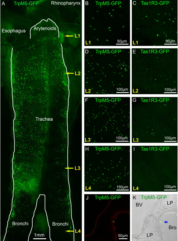
Background: Chemical irritation of airway mucosa elicits a variety of reflex responses such as coughing, apnea, and laryngeal closure. Inhaled irritants can activate either chemosensitive free nerve endings, laryngeal taste buds or solitary chemosensory cells (SCCs). The SCC population lies in the nasal respiratory epithelium, vomeronasal organ, and larynx, as well as deeper in the airway. The objective of this study is to map the distribution of SCCs within the airways and to determine the elements of the chemosensory transduction cascade expressed in these SCCs.

Methods: We utilized a combination of immunohistochemistry and molecular techniques (rtPCR and in situ hybridization) on rats and transgenic mice where the Tas1R3 or TRPM5 promoter drives expression of green fluorescent protein (GFP).

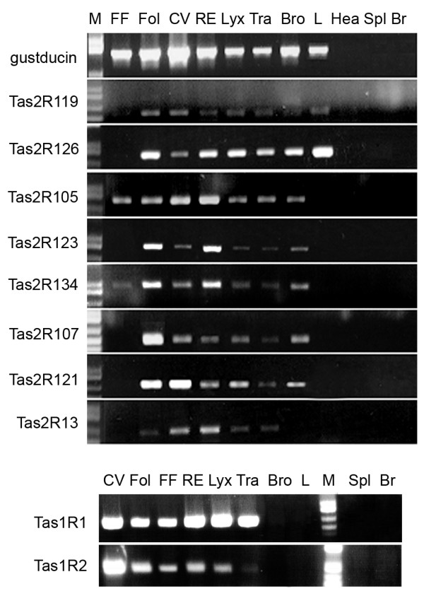
Results: Epithelial SCCs specialized for chemoreception are distributed throughout much of the respiratory tree of rodents. These cells express elements of the taste transduction cascade, including Tas1R and Tas2R receptor molecules, α-gustducin, PLCβ2 and TrpM5. The Tas2R bitter taste receptors are present throughout the entire respiratory tract. In contrast, the Tas1R sweet/umami taste receptors are expressed by numerous SCCs in the nasal cavity, but decrease in prevalence in the trachea, and are absent in the lower airways.

Conclusions: Elements of the taste transduction cascade including taste receptors are expressed by SCCs distributed throughout the airways. In the nasal cavity, SCCs, expressing Tas1R and Tas2R taste receptors, mediate detection of irritants and foreign substances which trigger trigeminally-mediated protective airway reflexes. Lower in the respiratory tract, similar chemosensory cells are not related to the trigeminal nerve but may still trigger local epithelial responses to irritants. In total, SCCs should be considered chemoreceptor cells that help in preventing damage to the respiratory tract caused by inhaled irritants and pathogens.


Figures

Figure 1
Figure 1
Morphological relationships of SCCs in the airways. A. Schematic representation of a sagittal section through the head of a mouse showing the airways and position of the different micrographs (green, blue and red rectangles). B & C. SCCs in the nasal respiratory epithelium. SCCs expressing TrpM5 or Tas1R3 (driven GFP) are intimately innervated by nerve fibers immunoreactive for PGP9.5 (B) or peptidergic nerve fibers of the trigeminal nerve immunoreactive for CGRP (C). D & E. In the larynx, Tas1R3-GFP+ SCCs are richly innervated by peptidergic (Substance P-immunoreactive) fibers probably from the superior laryngeal nerve (branch of the vagus) which shows responses to chemicals applied to the larynx [5,50]. F. SCCs of the trachea as shown by TRPM5-driven GFP are not closely embraced by nerve fibers although occasional en passant contacts can be observed.
Figure 2
Figure 2
SCCs are present to a different extent in the lower airways of TrpM5- and Tas1R3-GFP mice. A. Micrograph of whole-mounted larynx and trachea opened laterally to show the distribution of the SCC cells (exhibiting green TrpM5-driven GFP fluorescence). Some SCCs are present in the hypoglossal portion of the larynx just below the arytenoids. Numerous TrpM5 GFP+ SCCs are present throughout the length of the trachea although at a lower density in the bronchi. L1, L2, L3, L4 refer to the levels of the micrographs of panels B-K of this figure. L1 = hypoglottis, L2 = proximal trachea, L3 = distal trachea, L4 = bronchi. B-I. Whole mount en face views of epithelium from different levels of the trachea in the two transgenic lines, TrpM5-GFP and Tas1R3-GFP B. & C. SCCs in the hypoglossal region. D. - G. Numerous SCCs are present in the proximal (L2) and distal (L3) portions of the trachea in the TrpM5-GFP line, while fewer are evident in the Tas1R3-GFP line. H. & I. SCCs are still evident in the bronchi of the TrpM5-GFP mouse while virtually none express Tas1R3-driven GFP. J & K. Tissue section through the lung showing SCCs in bronchioles of more than 400 ± 100 μm in diameter (I), but none in smaller bronchioles or alveoli. J. Green channel fluorescent image. K. Identical image field showing a Normarski image along with the fluorescence image. An SCC is indicated by a blue arrow in panel K. BV = blood vessel, LP = lung parenchyma, Bro = bronchioles.
Figure 3
Figure 3
SCCs in the lower airways express elements of the taste transduction cascade. A-F. Single color channel (A & B; D & E) and merged (C & F) images of Tas1R3 GFP+ SCC cells in the trachea co-express TrpM5 and α-gustducin. G-L. Single color channel (G&H; J&K) and merged (I & L) images of TrpM5 GFP+ SCC cells in trachea (G-I) and bronchi (J-L) co-express α-gustducin. The insets of panels G-I show that α-gustducin is expressed only in a subset of the total TrpM5-GFP cell population (scale bar 10 μm for insets). M-O. Single color channel (M & N) and merged (O) images showing TrpM5 GFP+ SCC cells in the trachea co-express Plcβ2.
Figure 4
Figure 4
Rat airways express the Tas1R3 receptor and the G-protein α-gustducin. A. A PCR product for the α-gustducin gene is present in taste, respiratory and gastrointestinal tissue samples. B-G. Antisense probe (AS) for α-gustducin hybridizes with taste cells (B-D), as well as taste buds in the epiglottis (red arrows; F) and SCCs in the rhinopharynx and trachea (green arrows; E and G). H. The Tas1R3 gene PCR product is present in taste and airway tissue, but is not detectable in bronchi and lung. In contrast PCR for α-gustducin is positive in rat bronchi and lungs. Testis are positive for α-gustducin, but not for Tas1R3 I-N. The Tas1R3 AS probe hybridizes to taste tissue (I-K), laryngeal epithelium (red arrows indicate location of the taste buds; L-M) and a few SCCs in the trachea (green arrow; N). None of the negative control tissue (heart, spleen, liver and brain) showed expression of these two genes in PCR. Likewise, sense-control in situ probes showed no signal in any epithelium (Additional file 3). CV = circumvallate papillae; Fol = foliate papillae; Fung = fungiform papillae.
Figure 5
Figure 5
Rat airways express members of the Tas1R receptor family and several Tas2R bitter receptors. All of the Tas2R genes analyzed are expressed in the CV and Fol papillae, whereas only Tas2R105 and Tas2R134 are detected in the FF papillae. All tested Tas2Rs are detected in the nasal respiratory epithelium, larynx and trachea. With the exception of Tas2R13, all Tas2Rs are detected in the bronchi, but only Tas2R119 and Tas2R126 in the lung. None of the negative control tissue (heart, spleen and brain) showed any Tas2Rs or Tas1Rs expression. M = weight molecular marker; CV = circumvallate papillae; Fol = foliate papillae; FF = fungiform papillae; RE = respiratory epithelium; Lyx = larynx; Tra = trachea; Bro = bronchi; L = lung; Hea = heart; Spl = spleen; Br = brain.

References

    1. Widdicombe JG. Chemoreceptor control of the airways. Respir Physiol. 1992;87(3):373–381. doi: 10.1016/0034-5687(92)90018-R. - DOI - PubMed
    1. Bradley RM, Stedman HM, Mistretta CM. Superior laryngeal nerve response patterns to chemical stimulation of sheep epiglottis. Brain Res. 1983;276(1):81–93. doi: 10.1016/0006-8993(83)90550-4. - DOI - PubMed
    1. Stedman HM, Mistretta CM, Bradley RM. A quantitative study of cat epiglottal taste buds during development. J Anat. 1983;136(Pt 4):821–827. - PMC - PubMed
    1. Altschuler SM. Laryngeal and respiratory protective reflexes. Am J Med. 2001;111(Suppl 8A):90S–94S. doi: 10.1016/S0002-9343(01)00862-2. - DOI - PubMed
    1. Smith DV, Hanamori T. Organization of gustatory sensitivities in hamster superior laryngeal nerve fibers. J Neurophysiol. 1991;65(5):1098–1114. - PubMed

Publication types

MeSH terms