Review
. 2011 Apr;93(4):488-508.
doi: 10.1016/j.pneurobio.2011.01.004. Epub 2011 Jan 11.

Connecting the ear to the brain: Molecular mechanisms of auditory circuit assembly

Affiliations
Review

Connecting the ear to the brain: Molecular mechanisms of auditory circuit assembly

Jessica M Appler et al. Prog Neurobiol. 2011 Apr.

Abstract

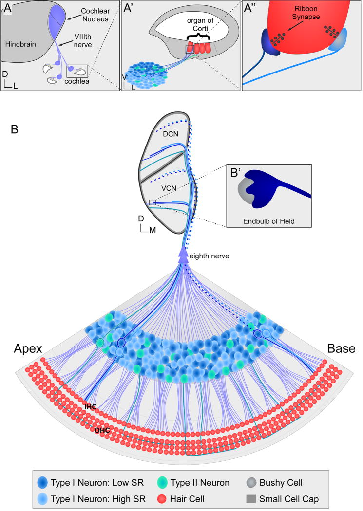
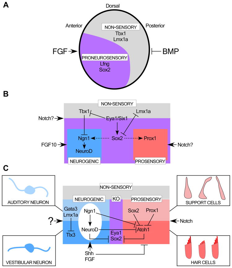
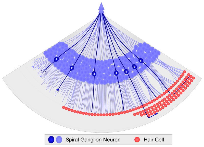
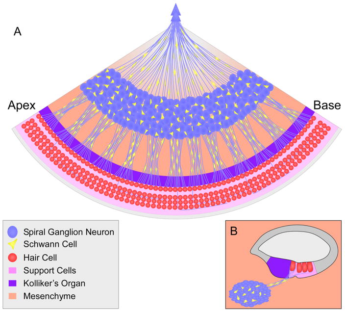
Our sense of hearing depends on precisely organized circuits that allow us to sense, perceive, and respond to complex sounds in our environment, from music and language to simple warning signals. Auditory processing begins in the cochlea of the inner ear, where sounds are detected by sensory hair cells and then transmitted to the central nervous system by spiral ganglion neurons, which faithfully preserve the frequency, intensity, and timing of each stimulus. During the assembly of auditory circuits, spiral ganglion neurons establish precise connections that link hair cells in the cochlea to target neurons in the auditory brainstem, develop specific firing properties, and elaborate unusual synapses both in the periphery and in the CNS. Understanding how spiral ganglion neurons acquire these unique properties is a key goal in auditory neuroscience, as these neurons represent the sole input of auditory information to the brain. In addition, the best currently available treatment for many forms of deafness is the cochlear implant, which compensates for lost hair cell function by directly stimulating the auditory nerve. Historically, studies of the auditory system have lagged behind other sensory systems due to the small size and inaccessibility of the inner ear. With the advent of new molecular genetic tools, this gap is narrowing. Here, we summarize recent insights into the cellular and molecular cues that guide the development of spiral ganglion neurons, from their origin in the proneurosensory domain of the otic vesicle to the formation of specialized synapses that ensure rapid and reliable transmission of sound information from the ear to the brain.


Figures

Figure 1
Figure 1. The organization of connections from the ear to the brain
(A) Diagrammatic view of a cross-section through the hindbrain at the level of the cochlea. Dorsal is up; lateral is right. Spiral ganglion neurons (blue) convey auditory information from the cochlea via the eighth (VIIIth) nerve, which arborizes within the cochlear nucleus in the hindbrain. A single turn of the cochlea is boxed and shown at higher power in A′. (A′) By convention, the orientation is flipped such that ventral is now up. Projections from spiral ganglion neurons (light blue, dark blue and green) penetrate the cochlear duct to reach the organ of Corti, which houses the hair cells (red). In this cross-sectional view, spiral ganglion neurons with a low spontaneous rate (SR) (dark blue neurons) are located more ventrally than those with high SR (light blue neurons). The boxed region is shown at higher power in A″. (A″) Spiral ganglion neurons receive information from hair cells via ribbon synapses. Connections made by low SR neurons (dark blue synapse) are located on the neural (also called the modiolar) side of the hair cell, while those made by high SR neurons (light blue synapse) are located on the on the abneural side. In this diagram, neural is to the left and abneural is to the right. (B) Schematic view of a wedge from a flatmounted cochlea (bottom) and its connections with the cochlear nucleus complex (top). In the cochlea, peripheral projections are corralled in radial bundles that pass through the spiral lamina to the hair cells (red). Low SR (dark blue) and high SR (light blue) Type I neurons contact inner hair cells (IHC). Type II neurons (green) are positioned in the ganglia nearest to the hair cells, and extend a projection past the inner hair cells and turn towards the base, with each projection contacting multiple outer hair cells (OHC) along its length. Information is conveyed to the cochlear nucleus by central axons, which bundle together to form the eighth nerve (double arrowhead). Upon entering the brainstem, individual axons bifurcate. The ascending projections terminate with bouton endings in the dorsal cochlear nucleus (DCN), while the descending projections target the ventral cochlear nucleus (VCN), where they form boutons with a variety of post-synaptic target neurons as well as unusual endbulb of Held synapses with bushy cells (B′). Within each division of the cochlear nucleus, auditory axons are tonotopically organized, such that high frequency information from the base of the cochlea is processed dorsally (dotted lines) and low frequency information from the apex is processed more ventrally (solid lines). In addition, the central projections from neurons with low spontaneous firing rates project more laterally than those with high spontaneous firing rates. Type II neurons project to the small cell cap that surrounds the cochlear nucleus complex (dark gray), as do some arbors from low SR fibers.
Figure 2
Figure 2. Parcellation of the otic vesicle
(A) Diagram of the early otic vesicle, as viewed laterally. The sensory epithelial cells and neurons develop within a proneurosensory domain (purple) in the anteroventral quadrant of the otic vesicle. This domain is marked by expression of Lfng and Sox2, while the complementary non-sensory domain expresses Tbx1 and Lmx1a. This initial patterning event may be controlled by FGF and BMP signals. (B) Summary of the transcriptional interactions that divide the non-sensory (gray), neurogenic (blue) and prosensory (red) lineages. Signals that are thought to mediate specific fate decisions are indicated outside the boxes. Possible interactions that require further confirmation are noted with dashed lines. Note that this diagram is highly schematic and meant to illustrate events that may occur even within a single cell as it selects its final fate. In reality, the neurogenic and prosensory domains are contained within the Sox2-positive proneurosensory domain, with a neurogenic domain in the center and multiple prosensory domains that are either contained within the neurogenic domain (i.e. for the utricle and saccule) or are on the periphery of the proneurosensory domain (i.e. for the cristae and cochlea). (C) The neurogenic domain ultimately produces either auditory or vestibular neurons, while cell-cell interactions within the prosensory domain create a mosaic of hair cells surrounded by support cells. Kolliker’s organ (KO) retains multipotent progenitors that share features with the initial progenitors in the proneurosensory domain.
Figure 3
Figure 3. The development of peripheral processes in the cochlea
Diagram of the cellular events that occur as spiral ganglion neurons (blue) develop in the cochlea, with immature cells shown on the left (1) and progressively maturing towards the right (5). Soon after becoming post-mitotic (1), the neurons become bipolar, with a central process extending into the eighth nerve and a peripheral process exploring the spiral lamina. Subsequently, neurons develop unbranched processes (2) that grow towards the edge of the organ of Corti, even before hair cells (red) have differentiated. Processes appear to wait while hair cell differentiation continues (3). Subsequently, Type I neurons (4) terminate beneath IHCs while Type II neurons (4′) develop large growth cones that navigate past the IHCs and into the rows of OHCs, where they turn towards the base, resulting in a mature pattern of innervation by birth (5, 5′).
Figure 4
Figure 4. The heterogeneous environment of the cochlea
A summary of the diverse cell types encountered by spiral ganglion neurons within the developing and mature cochlea, which is shown both as a flatmount (A) and in transverse section (B).

Similar articles

Cited by

References

    1. Abello G, Alsina B. Establishment of a proneural field in the inner ear. Int J Dev Biol. 2007;51:483–493. - PubMed
    1. Abello G, Khatri S, Radosevic M, Scotting PJ, Giraldez F, Alsina B. Independent regulation of Sox3 and Lmx1b by FGF and BMP signaling influences the neurogenic and non-neurogenic domains in the chick otic placode. Dev Biol. 2010;339:166–178. - PubMed
    1. Abraira VE, Del Rio T, Tucker AF, Slonimsky J, Keirnes HL, Goodrich LV. Cross-repressive interactions between Lrig3 and netrin 1 shape the architecture of the inner ear. Development. 2008;135:4091–4099. - PMC - PubMed
    1. Adam J, Myat A, Le Roux I, Eddison M, Henrique D, Ish-Horowicz D, Lewis J. Cell fate choices and the expression of Notch, Delta and Serrate homologues in the chick inner ear: parallels with Drosophila sense-organ development. Development. 1998;125:4645–4654. - PubMed
    1. Adamson CL, Reid MA, Davis RL. Opposite actions of brain-derived neurotrophic factor and neurotrophin-3 on firing features and ion channel composition of murine spiral ganglion neurons. J Neurosci. 2002;22:1385–1396. - PMC - PubMed

Publication types