Reduced somatostatin in subgenual anterior cingulate cortex in major depression
- PMID: 21232602
- PMCID: PMC3039077
- DOI: 10.1016/j.nbd.2011.01.014
Reduced somatostatin in subgenual anterior cingulate cortex in major depression
Abstract
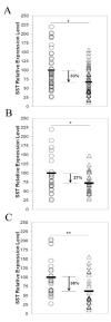
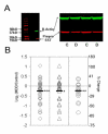
Converging evidence suggests a central role for dysfunction of the subgenual anterior cingulate cortex (sgACC) in the pathophysiology of major depressive disorder (MDD). Underlying mechanisms may include altered GABAergic function. Expression of somatostatin (SST), an inhibitory neuropeptide localized to a subset of GABA neurons, has been shown to be lower in the dorsolateral prefrontal cortex of male MDD subjects. Here, to investigate whether alterations in SST may contribute to sgACC dysfunction in MDD, and whether the alterations display sex-specificity, we measured sgACC SST at the mRNA and precursor peptide levels in a large cohort of subjects with MDD. SST mRNA levels were analyzed by quantitative PCR (qPCR) in the postmortem sgACC from male (n=26) and female (n=25) subjects with MDD and sex-matched subjects with no psychiatric diagnosis (n=51). Prepro-SST protein levels were assessed in a subset of subjects (n=42 pairs) by semi-quantitative Western blot. The mRNA expression of SST was significantly reduced by 38% in female subjects and by 27% in male subjects with MDD. The characteristic age-related decline in SST expression was observed in control (Pearson R=-0.357, p=0.005) but not MDD (R=-0.104, p=0.234) subjects, as low expression was detected across ages in MDD subjects. Protein expression was similarly reduced by 19% in both MDD groups, and findings were more robust in female (p=0.0056) than in males (p=0.0373) compared to respective controls. In conclusion, low SST represents a robust pathological finding in MDD. Specifically, alterations in SST signaling and/or SST-bearing GABA neurons may represent a critical pathophysiological entity that contributes to sgACC dysfunction and that matches to the high female vulnerability to develop MDD.
Copyright © 2010 Elsevier Inc. All rights reserved.
Figures


References
-
- Agid Y, Buzsaki G, Diamond DM, Frackowiak R, Giedd J, Girault JA, et al. How can drug discovery for psychiatric disorders be improved? Nat Rev Drug Discov. 2007;6:189–201. - PubMed
-
- Baraban SC, Tallent MK. Interneuron Diversity series: Interneuronal neuropeptides--endogenous regulators of neuronal excitability. Trends Neurosci. 2004;27:135–42. - PubMed
-
- Beasley CL, Zhang ZJ, Patten I, Reynolds GP. Selective deficits in prefrontal cortical GABAergic neurons in schizophrenia defined by the presence of calcium-binding proteins. Biol Psychiatry. 2002;52:708–715. - PubMed
-
- Cotter D, Landau S, Beasley C, Stevenson R, Chana G, MacMillan L, et al. The density and spatial distribution of GABAergic neurons, labelled using calcium binding proteins, in the anterior cingulate cortex in major depressive disorder, bipolar disorder, and schizophrenia. Biol Psychiatry. 2002;51:377–386. - PubMed
-
- Erraji-Benchekroun L, Underwood MD, Arango V, Galfalvy H, Pavlidis P, Smyrniotopoulos P, et al. Molecular aging in human prefrontal cortex is selective and continuous throughout adult life. Biol Psychiatry. 2005;57:549–58. - PubMed
Publication types
MeSH terms
Substances
Grants and funding
LinkOut - more resources
Full Text Sources

