. 2011 Apr 1;55(3):1034-43.
doi: 10.1016/j.neuroimage.2010.12.086. Epub 2011 Jan 11.

Data-driven optimization and evaluation of 2D EPI and 3D PRESTO for BOLD fMRI at 7 Tesla: I. Focal coverage

Affiliations

Data-driven optimization and evaluation of 2D EPI and 3D PRESTO for BOLD fMRI at 7 Tesla: I. Focal coverage

Robert L Barry et al. Neuroimage. .

Abstract

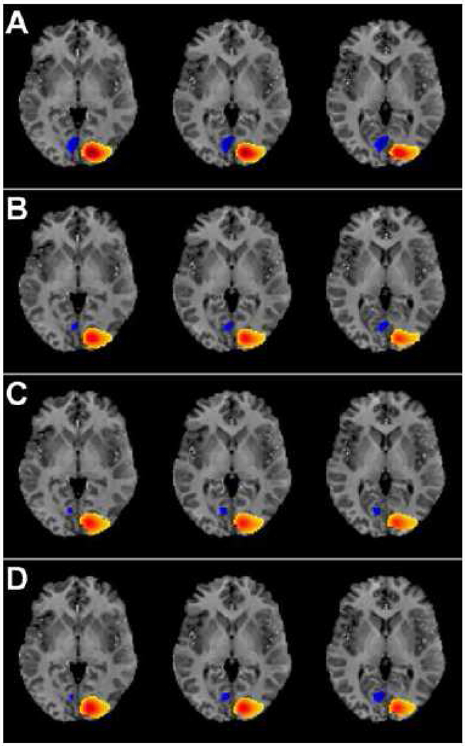
Blood oxygenation level dependent (BOLD) functional magnetic resonance imaging (fMRI) is commonly performed using 2D single-shot echo-planar imaging (EPI). However, single-shot EPI at 7 Tesla (T) often suffers from significant geometric distortions (due to low bandwidth (BW) in the phase-encode (PE) direction) and amplified physiological noise. Recent studies have suggested that 3D multi-shot sequences such as PRESTO may offer comparable BOLD contrast-to-noise ratio with increased volume coverage and decreased geometric distortions. Thus, a four-way group-level comparison was performed between 2D and 3D acquisition sequences at two in-plane resolutions. The quality of fMRI data was evaluated via metrics of prediction and reproducibility using NPAIRS (Non-parametric Prediction, Activation, Influence and Reproducibility re-Sampling). Group activation maps were optimized for each acquisition strategy by selecting the number of principal components that jointly maximized prediction and reproducibility, and showed good agreement in sensitivity and specificity for positive BOLD changes. High-resolution EPI exhibited the highest z-scores of the four acquisition sequences; however, it suffered from the lowest BW in the PE direction (resulting in the worst geometric distortions) and limited spatial coverage, and also caused some subject discomfort through peripheral nerve stimulation (PNS). In comparison, PRESTO also had high z-scores (higher than EPI for a matched in-plane resolution), the highest BW in the PE direction (producing images with superior geometric fidelity), the potential for whole-brain coverage, and no reported PNS. This study provides evidence to support the use of 3D multi-shot acquisition sequences in lieu of single-shot EPI for ultra high field BOLD fMRI at 7T.


Figures

FIG. 1
FIG. 1
Images acquired from a representative subject. (A) High-resolution anatomical corresponding to the middle functional slice (parallel to the calcarine sulcus). The rectangle depicts the approximate intersection of the shim volume through this slice. (B–E): The middle functional slice from the first volume acquired using (B) EPI with 1.19 × 1.19 × 2 mm3 voxels, (C) EPI with 2.19 × 2.19 × 2 mm3 voxels, (D) FFE with 1.19 × 1.19 × 2 mm3 voxels, and (E) PRESTO with 2.19 × 2.19 × 2 mm3 voxels. The outline of the brain (excluding dura) in (A) is superimposed on functional images to better visualize geometric distortions in the phase-encode direction (left to right) both within and outside the shim volume for each acquisition sequence.
FIG. 2
FIG. 2
(A–D): Plots of prediction vs. reproducibility (p,r) for images acquired using (A) EPI with 1.19 × 1.19 × 2 mm3 voxels, (B) EPI with 2.19 × 2.19 × 2 mm3 voxels, (C) FFE with 1.19 × 1.19 × 2 mm3 voxels, and (D) PRESTO with 2.19 × 2.19 × 2 mm3 voxels. All possible combinations of 7 spatial smoothing kernel sizes (7, 9, 11, 13, 15, 17, 19 mm FWHM; see legend in A) and 52 ranges of principal components (PCs) (1–2, 1–3, …, 1–49, 1–50, 1–75, 1–100, 1–150) are considered, resulting in 364 (p,r) pairs for each of the four plots. In theory, noiseless fMRI data with a perfect model would map to the point (1,1) in the top right corner. Concentric dotted curves mark points that are equidistant to (1,1), and the dashed line marks equal prediction and reproducibility (p = r). A very low number of PCs (1–2 or 1–3) can result in high reproducibility but low prediction from artifacts, illustrating why reproducibility alone is typically insufficient to identify the underlying dimensionality of the data. Increasing the number of PCs has a similar impact on prediction and reproducibility across kernel sizes and acquisition strategies: (p,r) increases toward (1,1) in a complicated manner, achieves one or more points that are close to (1,1), and then reproducibility decreases toward zero while prediction remains virtually unchanged. (E): For each of the 28 curves in (A–D), the (p,r) point with the shortest Euclidean distance to (1,1) is identified. (Az–Dz): The clusters outlined in (E) are magnified to better visualize the relative distance between points. The two numbers beside each point denote smoothing kernel size and number of PCs. The arrows and additional subplot labels (e.g., →3A) indicate which four (p,r) points correspond to the four activation maps in Fig. 3.
FIG. 3
FIG. 3
Group activation maps associated with optimal (p,r) (identified by arrows in Fig. 2(Az–Dz)) for: (A) high-resolution EPI with PCs=1–12, (B) low-resolution EPI with PCs=1–10, (C) FFE with PCs=1–10, and (D) PRESTO with PCs=1–13. Data were processed with a spatial smoothing kernel of 13 mm FWHM. Maps are presented with a common threshold of |z| > 5 and dynamic range from z = −6.670 (blue) to z = 18.99 (dark red).
FIG. 4
FIG. 4
Voxel-wise comparisons of z-scores for unthresholded activation maps in Fig. 3: (A) high-resolution (HR)-EPI vs. low-resolution (LR)-EPI; (B) HR-EPI vs. FFE; (C) HR-EPI vs. PRESTO; (D) FFE vs. LR-EPI; (E) PRESTO vs. LR-EPI; and (F) PRESTO vs. FFE. A point (representing one voxel) above the line of unity reflects a voxel exhibiting a higher group z-score with the acquisition sequence labeled on the vertical axis than the acquisition sequence labeled on the horizontal axis, and vice versa.

References

    1. Andersen AH, Gash DM, Avison MJ. Principal component analysis of the dynamic response measured by fMRI: a generalized linear systems framework. Magn. Reson. Imag. 1999;17:795–815. - PubMed
    1. Barry RL, Williams JM, Klassen LM, Gallivan JP, Culham JC, Menon RS. Evaluation of preprocessing steps to compensate for magnetic field distortions due to body movements in BOLD fMRI. Magn. Reson. Imag. 2010;28:235–244. - PMC - PubMed
    1. Bowen CV, Menon RS, Gati JS. High field balanced-SSFP fMRI: a BOLD technique with excellent tissue sensitivity and superior large vessel suppression. Proc. 13th Scientific Meeting of the ISMRM; Miami Beach, Florida. 2005. p. 119.
    1. Chen X, Pereira F, Lee W, Strother S, Mitchell T. Exploring predictive and reproducible modeling with the single-subject FIAC dataset. Hum. Brain Mapp. 2006;27:452–461. - PMC - PubMed
    1. Cohen MS, Weisskoff RM, Rzedzian RR, Kantor HL. Sensory stimulation by time-varying magnetic fields. Magn. Reson. Med. 1990;14:409–414. - PubMed

Publication types