

Molecular imaging in myeloma precursor disease
- PMID: 21232655
- PMCID: PMC3023940
- DOI: 10.1053/j.seminhematol.2010.11.006
Molecular imaging in myeloma precursor disease
Abstract
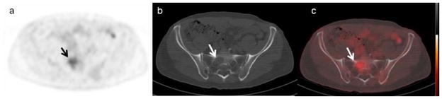
Multiple myeloma (MM) is consistently preceded by its pre-malignant states, monoclonal gammopathy of undetermined significance (MGUS) and/or smoldering multiple myeloma (SMM). By definition, precursor conditions do not exhibit end-organ disease (anemia, hypercalcemia, renal failure, skeletal lytic lesions, or a combination of these). However, new imaging methods are demonstrating that some patients in the MGUS or SMM category are exhibiting early signs of MM. Although MGUS/SMM patients are currently defined as low-risk versus high-risk based on clinical markers, we currently lack the ability to predict the individual patient's risk of progression from MGUS/SMM to MM. Given that the presence of gross lytic bone lesions is a hallmark of MM, it is reasonable to believe that less severe bone changes defined by more sensitive imaging may be predictive of MM progression. Indeed, since bone disease is such an essential aspect of MM, imaging techniques directed at the detection of early bone lesions, have the potential to become increasingly more useful in the setting of MGUS/SMM. Current guidelines for the radiological assessment of MM still recommend the traditional skeletal survey, although its limitations are well documented, especially in early phases of the disease when radiographs can significantly underestimate the extent of bone lesions and bone marrow involvement. Newer, more advanced imaging modalities, with higher sensitivities, including whole-body low-dose computed tomography (CT), magnetic resonance imaging (MRI), and positron emission tomography (PET) are being employed. Also various imaging techniques have been used to provide an assessment of bone involvement and identify extra-osseous disease. This review emphasizes the current state of the art and emerging imaging methods, which may help to better define high-risk versus low-risk MGUS/SMM. Ultimately, improved imaging could allow more tailored clinical management, and, most likely play an important role in the development of future treatment strategies for high-risk precursor disease.
Published by Elsevier Inc.
Figures
Similar articles
-
Development of early treatment strategies for high-risk myeloma precursor disease in the future.Semin Hematol. 2011 Jan;48(1):66-72. doi: 10.1053/j.seminhematol.2010.11.009. Semin Hematol. 2011. PMID: 21232660 Free PMC article. Review.
-
Bone marrow abnormalities and early bone lesions in multiple myeloma and its precursor disease: a prospective study using functional and morphologic imaging.Leuk Lymphoma. 2016 May;57(5):1114-21. doi: 10.3109/10428194.2015.1090572. Epub 2016 Apr 7. Leuk Lymphoma. 2016. PMID: 26690712 Free PMC article.
-
[Imaging in smoldering (asymptomatic) multiple myeloma. Past, present and future].Radiologe. 2014 Jun;54(6):572, 574-81. doi: 10.1007/s00117-014-2694-7. Radiologe. 2014. PMID: 24927659 German.
-
Diagnosis, risk stratification and management of monoclonal gammopathy of undetermined significance and smoldering multiple myeloma.Int J Lab Hematol. 2016 May;38 Suppl 1:110-22. doi: 10.1111/ijlh.12504. Epub 2016 May 9. Int J Lab Hematol. 2016. PMID: 27161311 Review.
-
Development of a Multivariable Model to Predict the Need for Bone Marrow Sampling in Persons With Monoclonal Gammopathy of Undetermined Significance : A Cohort Study Nested in a Clinical Trial.Ann Intern Med. 2024 Apr;177(4):449-457. doi: 10.7326/M23-2540. Epub 2024 Apr 2. Ann Intern Med. 2024. PMID: 38560901
Cited by
-
Does my patient with a serum monoclonal spike have multiple myeloma?Hematol Oncol Clin North Am. 2012 Apr;26(2):383-93, ix. doi: 10.1016/j.hoc.2012.02.009. Hematol Oncol Clin North Am. 2012. PMID: 22463833 Free PMC article. Review.
-
Diagnostic and prognostic utility of non-invasive imaging in diabetes management.World J Diabetes. 2015 Jun 25;6(6):792-806. doi: 10.4239/wjd.v6.i6.792. World J Diabetes. 2015. PMID: 26131322 Free PMC article. Review.
-
MRI in multiple myeloma: a pictorial review of diagnostic and post-treatment findings.Insights Imaging. 2016 Aug;7(4):553-69. doi: 10.1007/s13244-016-0492-7. Epub 2016 May 10. Insights Imaging. 2016. PMID: 27164915 Free PMC article. Review.
-
Current Status and Future of Artificial Intelligence in MM Imaging: A Systematic Review.Diagnostics (Basel). 2023 Nov 2;13(21):3372. doi: 10.3390/diagnostics13213372. Diagnostics (Basel). 2023. PMID: 37958267 Free PMC article. Review.
-
Diagnostic performance of 18 F-FDG-PET/CT compared to standard skeletal survey for detecting bone destruction in smouldering multiple myeloma: time to move forward.Br J Haematol. 2021 Apr;193(1):125-128. doi: 10.1111/bjh.17088. Epub 2020 Sep 23. Br J Haematol. 2021. PMID: 32966607 Free PMC article.
References
-
- Landgren O, Kyle RA, Pfeiffer RM, Katzmann JA, Caporaso NE, Hayes RB, Dispenzieri A, Kumar S, Clark RJ, Baris D, Hoover R, Rajkumar SV. Monoclonal gammopathy of undetermined significance (MGUS) consistently precedes multiple myeloma: a prospective study. Blood. 2009 May 28;113(22):5412–7. - PMC - PubMed
-
- Kyle RA, Durie BG, Rajkumar SV, Landgren O, Blade J, Merlini G, et al. Monoclonal gammopathy of undetermined significance (MGUS) and smoldering (asymptomatic) multiple myeloma: IMWG consensus perspectives risk factors for progression and guidelines for monitoring and management. Leukemia. 2010 Jun;24(6):1121–7. - PMC - PubMed
-
- Kyle RA, Rajkumar SV. Multiple myeloma. N Engl J Med. 2004;351:1860–1873. - PubMed
-
- Rajkumar SV, Kyle RA. Multiple myeloma: diagnosis and treatment. Mayo Clin Proc. 2005;80:1371–1382. - PubMed
-
- Durie BGM, Salmon SE. A clinical staging system for multiple myeloma. Cancer. 1975;36:842–54. - PubMed
Publication types
MeSH terms
Grants and funding
LinkOut - more resources
Full Text Sources
Medical
