. 2011 Jan;4(1):1-10.
doi: 10.1016/j.jcmg.2010.06.018.

Adenine nucleotide translocase 1 deficiency results in dilated cardiomyopathy with defects in myocardial mechanics, histopathological alterations, and activation of apoptosis

Affiliations

Adenine nucleotide translocase 1 deficiency results in dilated cardiomyopathy with defects in myocardial mechanics, histopathological alterations, and activation of apoptosis

Nupoor Narula et al. JACC Cardiovasc Imaging. 2011 Jan.

Abstract

Objectives: the aim of this study was to test the hypothesis that chronic mitochondrial energy deficiency causes dilated cardiomyopathy, we characterized the hearts of age-matched young and old adenine nucleotide translocator (ANT)1 mutant and control mice.

Background: ANTs export mitochondrial adenosine triphosphate into the cytosol and have a role in the regulation of the intrinsic apoptosis pathway. Mitochondrial energy deficiency has been hypothesized, on the basis of indirect evidence, to be a factor in the pathophysiology of dilated cardiomyopathies. Ant1 inactivation should limit adenosine triphosphate for contraction and calcium transport, thereby resulting in early cardiac dysfunction with later dilation and heart failure.

Methods: we conducted a multiyear study of 73 mutant (Ant1-/-) and 57 control (Ant1+/+) mice, between the ages of 2 and 21 months. Hearts were characterized by cardiac anatomy, echocardiographic imaging with velocity vector analysis, histopathology, and apoptosis assays.

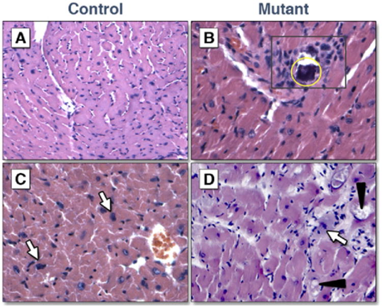
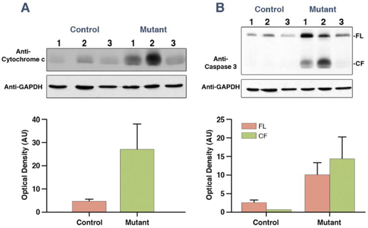
Results: the Ant1-/- mice developed a distinctive concentric dilated cardiomyopathy, characterized by substantial myocardial hypertrophy and ventricular dilation, with cardiac function declining earlier in age as compared to control mice. Left ventricular circumferential, radial, and rotational mechanics were reduced even in the younger mutants with preserved systolic function. Histopathologic analysis demonstrated increased myocyte hypertrophy, fibrosis, and calcification in the mutant mice as compared with control mice. Furthermore, increased cytoplasmic cytochrome c levels and caspase 3 activation were observed in the mutant mice.

Conclusions: our results demonstrate that mitochondrial energy deficiency is sufficient to cause dilated cardiomyopathy, confirming that energy defects are a factor in this disease. Energy deficiency initially leads to early mechanical dysfunction before a decline in left ventricular systolic function. Chronic energy deficiency with age then leads to heart failure. Our results now allow us to use the Ant1-/- mouse model for testing new therapies for ANT1 mutant patients.


Figures

Figure 1
Figure 1. Cardiomyopathy in Ant1−/− Mouse
(A) M-mode echocardiography. Ultrasound images depict left ventricular dimensions in a control (upper panel) and mutant (bottom panel) mouse. Interventricular septum (IVS) thickness is shown as distance between blue and red lines, and left ventricular internal dimension at end-diastole (LVIDd) is shown as distance between red and yellow lines. The LVIDd is increased from 3.3 mm in control to 3.9 mm in mutant. (B) Velocity vector imaging: radial and rotational velocity vectors. Left ventricular short axis in control (ac) and Ant1 mutant (df). Endocardial velocity vectors (a and d) were attenuated at end-ejection in mutant. The 2-dimensional maps of radial velocity and the apical rotational velocity are presented in adjacent panels. In contrast to the uniform pattern of radial velocities seen in control mice (b), marked dyssynchrony is seen in the mutant animal (e). Similarly, the velocity of counterclockwise rotation in systole is biphasic in the control mice (c) but is dyssynchronous in the mutant animal (f). (C) Velocity vector imaging: velocity, strain, and strain rates. Images from control (upper panels) and mutant (bottom panels). For each heart, velocity (a and d), strain (b and e), and strain rates (c and f) are depicted by 3-dimensional color mapping. Uniform motion and shortening are observed in control, whereas dyssynchronous motion and shortening are delineated in the velocity, strain, and strain rate curves of mutant.
Figure 2
Figure 2. EF Scatter Plot for Ant1 Mutants and Control Mice
Mutants (X) and control mice (circle). Abnormal ejection fraction (EF) is defined as >2 SD below the average EF of control animals (dashed horizontal line). The best-fit linear regression line is provided with R2 for each group. Sac = sacrificed.
Figure 3
Figure 3. Histopathological Alterations in Mutant Animals
(A) Control, 7.5-month-old, with mild hypertrophy; (B) mutant, 9-month-old, shows inflammation (boxed area) and calcification (circled area); (C) control, 17-month-old, shows normal myocardial architecture with mild hypertrophy (white arrows); (D) mutant, 18-month-old, shows basophilic degeneration (white arrow) and myofibrillar lysis (triangles).
Figure 4
Figure 4. Quantification of Histopathologic Features
(A and B) Hematoxylin and eosin stained; (C and D) Masson's trichrome stained; (A and C) aggregate of data from all ages; (B) aggregate of data from mice <12 months old; (D) aggregate of data from mice ≥12 months old. Binucl = binucleation; Calc = calcification; F = focal; Hyp = hypertrophy; Infl = inflammation; Int = interstitial; M = multifocal; MFL = myofibrillar lysis; Per = perivascular; Rep = replacement.
Figure 5
Figure 5. Cardiac Fibrosis by Masson's Trichrome Staining
(A) Control, 8.5-month-old, shows multifocal interstitial fibrosis; (B) mutant, 5.5-month-old, shows multifocal replacement fibrosis; (C) control, 14.5-month-old, shows no fibrosis; (D) mutant, 13.5-month-old, shows multifocal interstitial fibrosis; (E) control, 19-month-old, shows focal perivascular fibrosis and focal interstitial fibrosis (not seen here); (F) mutant, 18.5-month-old, shows focal replacement fibrosis and multiple foci of interstitial fibrosis.
Figure 6
Figure 6. Apoptosis Assays by Western Blot
(A) Increased cytochrome c levels in 3 mutants versus 3 control mice, ages >12 months, p = 0.05, error bars show SEM. Cytochrome c = 14 kDa, glyceraldehyde-3-phosphate dehydrogenase (GAPDH) = 38 kDa. (B) Increased inactive and active caspase 3 levels in mutants, error bars are SEM. Inactive caspase 3 = 32 kDa, active caspase 3 = 17 kDa. AU = arbitrary unit; CF = cleaved form; FL = full length; kDa = kilodaltons; SEM = standard error of the mean.

Comment in

References

    1. Portman MA. The adenine nucleotide translocator: regulation and function during myocardial development and hypertrophy. Clin Exp Pharmacol. 2002;29:334–8. - PubMed
    1. Pebay-Peyroula E, Dahout-Gonzalez C, Kahn R, et al. Structure of mitochondrial ADP/ATP carrier in complex with carboxyatractyloside. Nature. 2003;426:39–44. - PubMed
    1. Kokoszka JE, Waymire KG, Levy SE, et al. The ADP/ATP translocator is not essential for the mitochondrial permeability transition pore. Nature. 2004;427:461–5. - PMC - PubMed
    1. Bauer MK, Schubert A, Rocks O, Grimm S. Adenine nucleotide translocase-1, a component of the permeability transition pore, can dominantly induce apoptosis. J Cell Biol. 1999;147:1493–502. - PMC - PubMed
    1. Zamora M, Granell M, Mampel T, Vinas O. Adenine nucleotide translocase 3 (ANT3) overexpression induces apoptosis in cultured cells. FEBS Lett. 2004;563:155–60. - PubMed

Publication types

MeSH terms

Substances