

Review
doi: 10.1016/j.pain.2010.11.005.
Epub 2011 Jan 12.
Pathophysiology of postoperative pain
Affiliations
- PMID: 21232860
- PMCID: PMC3073562
- DOI: 10.1016/j.pain.2010.11.005
Item in Clipboard
Review
Pathophysiology of postoperative pain
Pain.
2011 Mar.
No abstract available
Conflict of interest statement
The author has no conflict of interest to declare.
Figures
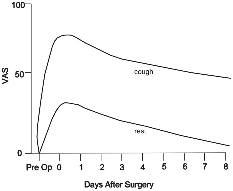
Schematic of postoperative pain after major surgery in patients with optimized parenteral opioid analgesia. Top line is pain with cough; bottom line is pain at rest. VAS=visual analogue scale.
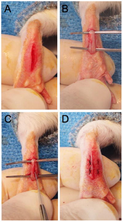
Photographs of the different stages of the rat plantar incision. A: A one cm longitudinal incision is made through the skin and fascia starting 0.5 cm from the proximal edge of the heel and extending toward the distal aspect of the paw. B–C: The underlying flexor muscle is elevated and also incised longitudinally. The muscle is split and dissected longitudinally. D: After hemostasis, the wound is apposed with two mattress sutures of 5-0 nylon.
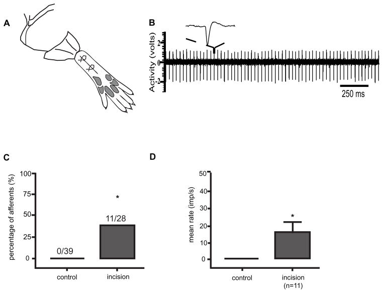
Spontaneous activity in nociceptors one day after plantar incision [29]. A: Schematic of in vivo recording from nociceptors from rats that underwent hindpaw incision. B: Example of spontaneous action potentials recorded from nociceptor in rat that underwent plantar incision. C: Percentage of nociceptors with spontaneous activity in the control group that underwent a sham operation and group that underwent skin, fascia and muscle incision [29; 42]. D: Mean rate of spontaneous activity of nociceptors that underwent skin, fascia and muscle incision [29; 42].
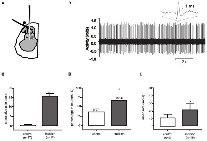
Spontaneous activity in dorsal horn neurons one day after plantar incision [43]. A: Schematic of dorsal horn neuron recording. B: Example of spontaneous action potentials recorded from dorsal horn neuron in a rat that underwent plantar incision and reproduced from Pain 144: 329–339 with permission of the International Association for the Study of Pain® (IASP®). The figure may not be reproduced for any other purpose without permission.. C: Guarding pain score in rats that underwent a sham (control) operation and group that underwent skin, fascia and muscle incision. D: Percentage of dorsal horn neurons with spontaneous activity in the control group and group that underwent skin, fascia and muscle incision. E: Mean rate of spontaneous activity of dorsal horn neurons in the control and incision groups. C–D adapted from [43] European Journal of Pain, Vol 13, Issue 8, Xu J, Richebe P, Brennan TJ, Separate groups of dorsal horn neurons transmit spontaneous activity and mechanosensitivity one day after plantar incision, p. 823 and 824, Copyright 2010, with permission from Elsevier.
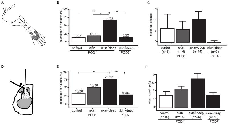
Spontaneous activity in nociceptors and dorsal horn neurons. A–C. Nociceptor recordings in rats that underwent a sham (control) operation, a skin incision, a skin, fascia and muscle incision and a group 7 days after skin, fascia and muscle incision. Percentage of nociceptors with spontaneous activity (B) and average spontaneous activity (C). B–C from [42]. Figure B–C from Xu J, Brennan TJ. Guarding pain and spontaneous activity of nociceptors after skin versus skin plus deep tissue incision. Anesthesiology 2010; 112:153–64 with permission from Lippincott, Williams, & Wilkins. D–F. Dorsal horn neuron recordings in rats that underwent a sham (control) operation, a skin incision, a skin, fascia and muscle incision and a group 7 days after skin, fascia and muscle incision. Percentage of dorsal horn neurons with spontaneous activity (E) and average spontaneous activity (F). E–F adapted from [41]. Figure E–F has been reproduced with permission of the International Association for the Study of Pain® (IASP®). The figure may not be reproduced for any other purpose without permission.
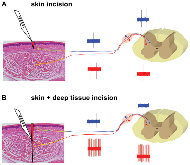
Schematic for the development guarding pain and SA in the nociceptive pathways after plantar incision. (A) An incision in the skin only (epidermis and dermis layer) induces minimal SA in nociceptors and DHNs which receive predominately cutaneous input. (B) An incision including skin and deep tissue (fascia and muscle layer) results in sustained SA in muscle-innervating primary afferents and the DHNs receiving input from muscle. From Xu J, Brennan TJ. Guarding pain and spontaneous activity of nociceptors after skin versus skin plus deep tissue incision. Anesthesiology 2010; 112:153–64 with permission from Lippincott, Williams, & Wilkins.
References
-
- Apfelbaum JL, Chen C, Mehta SS, Gan TJ. Postoperative pain experience: results from a national survey suggest postoperative pain continues to be undermanaged. Anesth Analg. 2003;97:534–540. table of contents. - PubMed
-
- Beitz AJ, Newman A, Shepard M, Ruggles T, Eikmeier L. A new rodent model of hind limb penetrating wound injury characterized by continuous primary and secondary hyperalgesia. J Pain. 2004;5:26–37. - PubMed
-
- Bessou P, Perl ER. Response of cutaneous sensory units with unmyelinated fibers to noxious stimuli. J Neurophysiol. 1969;32:1025–1043. - PubMed
-
- Brennan TJ, Kehlet H. Preventive analgesia to reduce wound hyperalgesia and persistent postsurgical pain: not an easy path. Anesthesiology. 2005;103:681–683. - PubMed
-
- Brennan TJ, Vandermeulen EP, Gebhart GF. Characterization of a rat model of incisional pain. Pain. 1996;64:493–501. - PubMed
Publication types
MeSH terms
Grants and funding
LinkOut - more resources
Full Text Sources
Other Literature Sources
