

A conditional Granger causality model approach for group analysis in functional magnetic resonance imaging
- PMID: 21232892
- PMCID: PMC3063394
- DOI: 10.1016/j.mri.2010.10.008
A conditional Granger causality model approach for group analysis in functional magnetic resonance imaging
Abstract
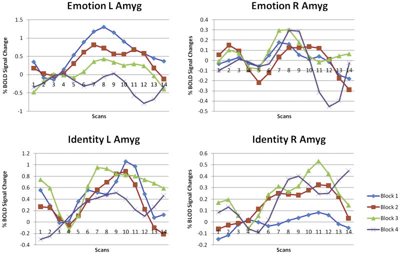
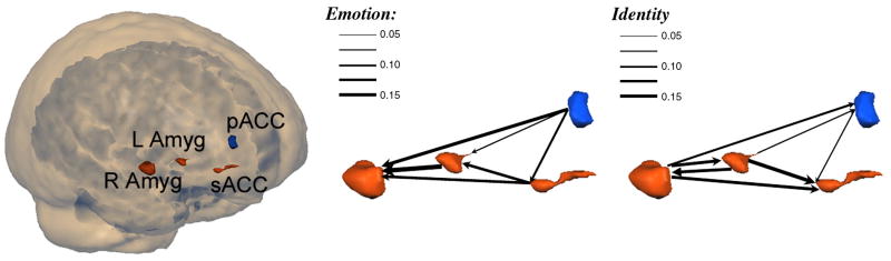
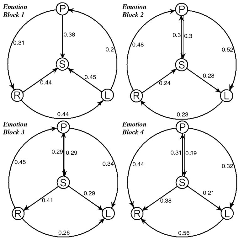
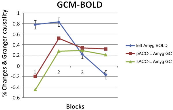
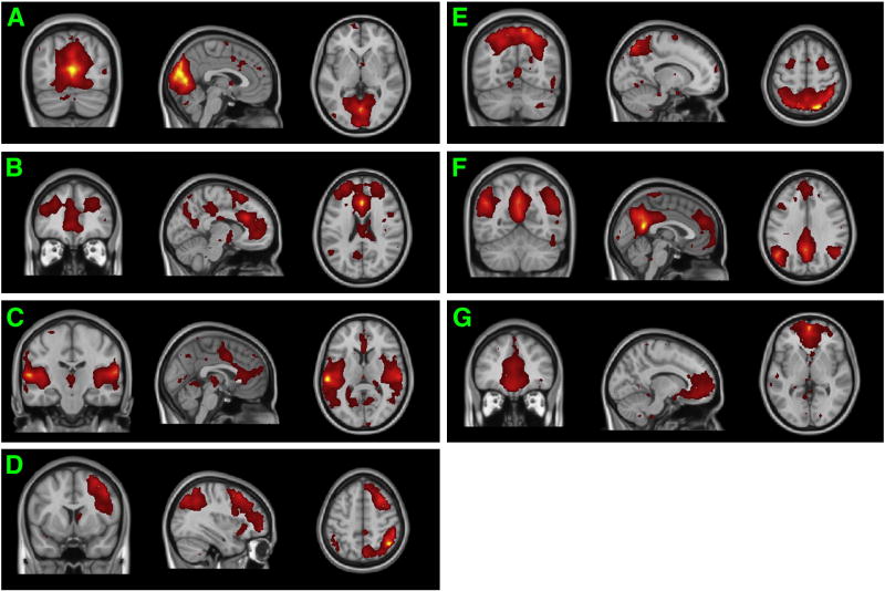
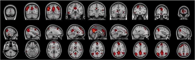
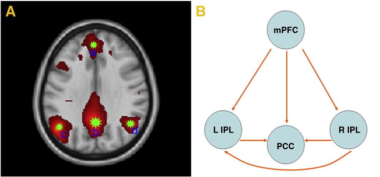
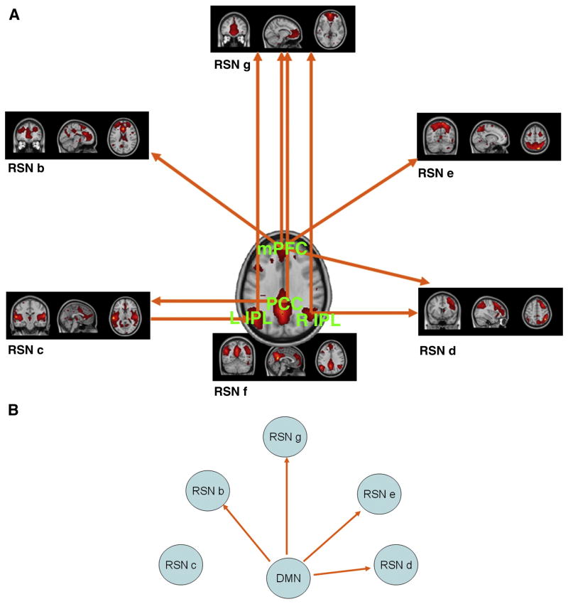
Granger causality model (GCM) derived from multivariate vector autoregressive models of data has been employed to identify effective connectivity in the human brain with functional magnetic resonance imaging (fMRI) and to reveal complex temporal and spatial dynamics underlying a variety of cognitive processes. In the most recent fMRI effective connectivity measures, pair-wise GCM has commonly been applied based on single-voxel values or average values from special brain areas at the group level. Although a few novel conditional GCM methods have been proposed to quantify the connections between brain areas, our study is the first to propose a viable standardized approach for group analysis of fMRI data with GCM. To compare the effectiveness of our approach with traditional pair-wise GCM models, we applied a well-established conditional GCM to preselected time series of brain regions resulting from general linear model (GLM) and group spatial kernel independent component analysis of an fMRI data set in the temporal domain. Data sets consisting of one task-related and one resting-state fMRI were used to investigate connections among brain areas with the conditional GCM method. With the GLM-detected brain activation regions in the emotion-related cortex during the block design paradigm, the conditional GCM method was proposed to study the causality of the habituation between the left amygdala and pregenual cingulate cortex during emotion processing. For the resting-state data set, it is possible to calculate not only the effective connectivity between networks but also the heterogeneity within a single network. Our results have further shown a particular interacting pattern of default mode network that can be characterized as both afferent and efferent influences on the medial prefrontal cortex and posterior cingulate cortex. These results suggest that the conditional GCM approach based on a linear multivariate vector autoregressive model can achieve greater accuracy in detecting network connectivity than the widely used pair-wise GCM, and this group analysis methodology can be quite useful to extend the information obtainable in fMRI.
Copyright © 2011 Elsevier Inc. All rights reserved.
Figures
Similar articles
-
Dynamic Granger causality based on Kalman filter for evaluation of functional network connectivity in fMRI data.Neuroimage. 2010 Oct 15;53(1):65-77. doi: 10.1016/j.neuroimage.2010.05.063. Epub 2010 Jun 1. Neuroimage. 2010. PMID: 20561919 Free PMC article.
-
Analyzing brain networks with PCA and conditional Granger causality.Hum Brain Mapp. 2009 Jul;30(7):2197-206. doi: 10.1002/hbm.20661. Hum Brain Mapp. 2009. PMID: 18830956 Free PMC article.
-
Analyzing the connectivity between regions of interest: an approach based on cluster Granger causality for fMRI data analysis.Neuroimage. 2010 Oct 1;52(4):1444-55. doi: 10.1016/j.neuroimage.2010.05.022. Epub 2010 Jun 1. Neuroimage. 2010. PMID: 20472076
-
Identifying the default mode network structure using dynamic causal modeling on resting-state functional magnetic resonance imaging.Neuroimage. 2014 Feb 1;86:53-9. doi: 10.1016/j.neuroimage.2013.07.071. Epub 2013 Aug 6. Neuroimage. 2014. PMID: 23927904 Free PMC article.
-
Assessing functional connectivity in the human brain by fMRI.Magn Reson Imaging. 2007 Dec;25(10):1347-57. doi: 10.1016/j.mri.2007.03.007. Epub 2007 May 11. Magn Reson Imaging. 2007. PMID: 17499467 Free PMC article. Review.
Cited by
-
Reduced Dynamic Interactions Within Intrinsic Functional Brain Networks in Early Blind Patients.Front Neurosci. 2019 Mar 22;13:268. doi: 10.3389/fnins.2019.00268. eCollection 2019. Front Neurosci. 2019. PMID: 30983956 Free PMC article.
-
Reentrant Information Flow in Electrophysiological Rat Default Mode Network.Front Neurosci. 2017 Feb 27;11:93. doi: 10.3389/fnins.2017.00093. eCollection 2017. Front Neurosci. 2017. PMID: 28289373 Free PMC article.
-
Altered effective connectivity in Parkinson's disease patients with rapid eye movement sleep behavior disorder: a resting-state functional magnetic resonance imaging study and support vector machine analysis.Quant Imaging Med Surg. 2025 Jan 2;15(1):352-369. doi: 10.21037/qims-24-1196. Epub 2024 Dec 27. Quant Imaging Med Surg. 2025. PMID: 39839023 Free PMC article.
-
Construct validation of a DCM for resting state fMRI.Neuroimage. 2015 Feb 1;106:1-14. doi: 10.1016/j.neuroimage.2014.11.027. Epub 2014 Nov 21. Neuroimage. 2015. PMID: 25463471 Free PMC article.
-
Granger causality analysis reveals distinct spatio-temporal connectivity patterns in motor and perceptual visuo-spatial working memory.Front Comput Neurosci. 2014 Nov 13;8:146. doi: 10.3389/fncom.2014.00146. eCollection 2014. Front Comput Neurosci. 2014. PMID: 25431557 Free PMC article.
References
-
- Abler B, Roebroeck A, Goebel R, Höse A, Schönfeldt-Lecuona C, Hole G, Walter H. Investigating directed influences between activated brain areas in a motor-response task using fMRI. Magn Reson Imaging. 2006;24:181–185. - PubMed
-
- Bach FR, Jordan M. Kernel Independent Component Analysis. Journal of Machine Learning Research. 2002:1–48.
-
- Brannan SK, Mayberg HS, McGinnis S. Cingulate metabolism predicts treatment response: a replication. Biol Psychiatry. 2000;47:107S.
-
- Buckner RL, Andrews-Hanna JR, Schacter DL. The brain's default network: anatomy, function, and relevance to disease. Ann N Y Acad Sci. 2008;1124:1–38. - PubMed
-
- Buckner RL, Carroll DC. Self-projection and the brain. Trends Cogn Sci. 2007;11:49–57. - PubMed
Publication types
MeSH terms
Grants and funding
LinkOut - more resources
Full Text Sources
Medical
