. 2011 Feb;52(2):188-95.
doi: 10.2967/jnumed.110.081463. Epub 2011 Jan 13.

Evaluation of 18F-FDG PET and MRI associations in pediatric diffuse intrinsic brain stem glioma: a report from the Pediatric Brain Tumor Consortium

Affiliations

Evaluation of 18F-FDG PET and MRI associations in pediatric diffuse intrinsic brain stem glioma: a report from the Pediatric Brain Tumor Consortium

Katherine A Zukotynski et al. J Nucl Med. 2011 Feb.

Abstract

The purpose of this study was to assess (18)F-FDG uptake in children with a newly diagnosed diffuse intrinsic brain stem glioma (BSG) and to investigate associations with progression-free survival (PFS), overall survival (OS), and MRI indices.

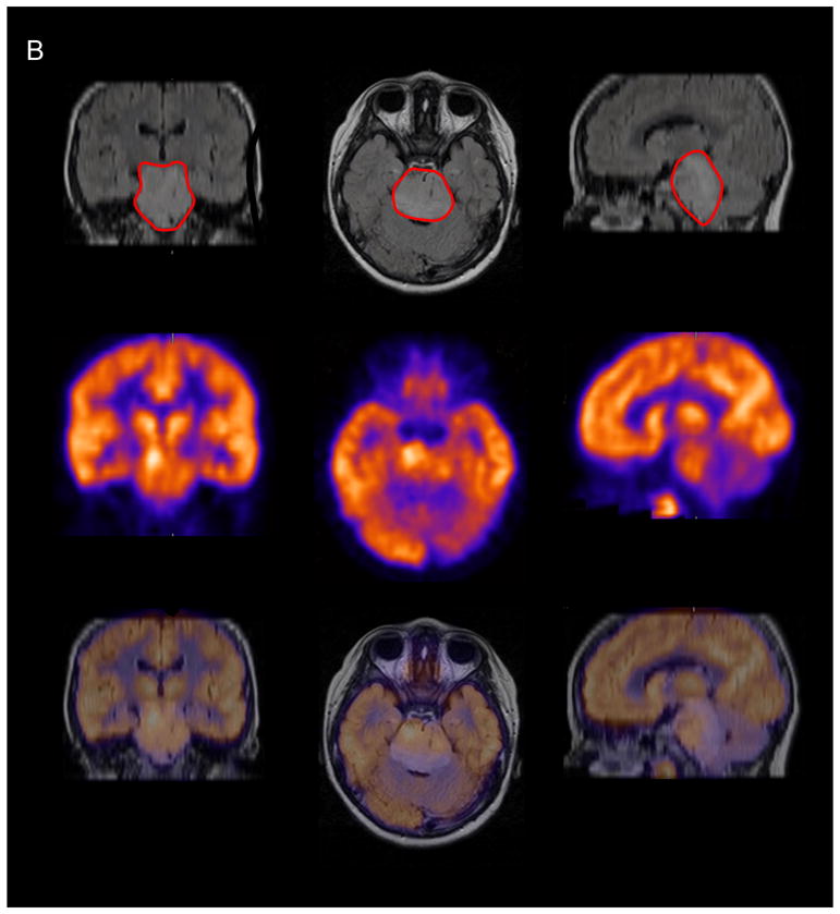
Methods: Two Pediatric Brain Tumor Consortium (PBTC) therapeutic trials in children with newly diagnosed BSG were designed to test radiation therapy combined with molecularly targeted agents (PBTC-007: phase I/II study of gefitinib; PBTC-014: phase I/II study of tipifarnib). Baseline brain (18)F-FDG PET scans were obtained in 40 children in these trials. Images were evaluated by consensus between 2 PET experts for intensity and uniformity of tracer uptake. Associations of (18)F-FDG uptake intensity and uniformity with both PFS and OS, as well as associations with tumor MRI indices at baseline (tumor volume on fluid-attenuated inversion recovery, baseline intratumoral enhancement, diffusion and perfusion values), were evaluated.

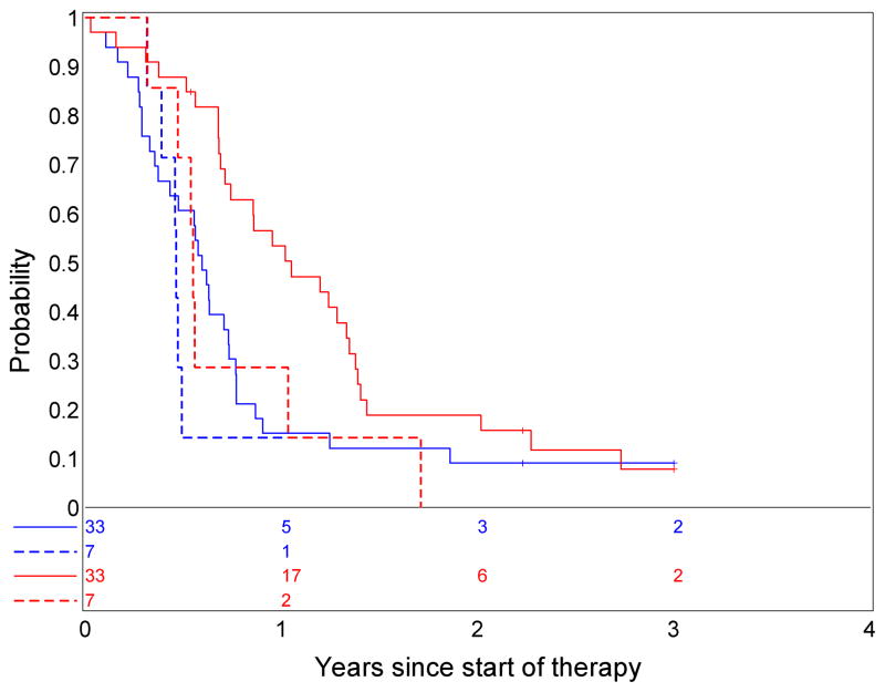
Results: In most of the children, BSG (18)F-FDG uptake was less than gray-matter uptake. Survival was poor, irrespective of intensity of (18)F-FDG uptake, with no association between intensity of (18)F-FDG uptake and PFS or OS. However, hyperintense (18)F-FDG uptake in the tumor, compared with gray matter, suggested poorer survival rates. Patients with (18)F-FDG uptake in 50% or more of the tumor had shorter PFS and OS than did patients with (18)F-FDG uptake in less than 50% of the tumor. There was some evidence that tumors with higher (18)F-FDG uptake were more likely to show enhancement, and when the diffusion ratio was lower, the uniformity of (18)F-FDG uptake appeared higher.

Conclusion: Children with BSG for which (18)F-FDG uptake involves at least half the tumor appear to have poorer survival than children with uptake in less than 50% of the tumor. A larger independent study is needed to verify this hypothesis. Intense tracer uptake in the tumors, compared with gray matter, suggests decreased survival. Higher (18)F-FDG uptake within the tumor was associated with enhancement on MR images. Increased tumor cellularity as reflected by restricted MRI diffusion may be associated with increased (18)F-FDG uniformity throughout the tumor.


Figures

FIGURE 1
FIGURE 1
Illustrative examples of the grading scheme for intensity and uniformity of 18F-labeled 2-fluoro-2-deoxy-D-glucose (18F-FDG) PET uptake by brainstem glioma. (A) 12-year-old girl with intensity of brainstem glioma 18F-FDG uptake between normal white and gray matter, the percentage of the tumor demonstrating 18F-FDG uptake <25%(as demonstrated on the FLAIR MRI-marked with red ROI), progression-free survival of 258 days and overall survival of 348 days. (B) 7-year-old boy with intensity of brainstem glioma 18F-FDG uptake greater than normal gray matter, the percentage of the tumor demonstrating 18F-FDG uptake >75% (as demonstrated on the FLAIR MRI marked with red ROI), progression-free survival of 169 days and overall survival of 196 days.
FIGURE 1
FIGURE 1
Illustrative examples of the grading scheme for intensity and uniformity of 18F-labeled 2-fluoro-2-deoxy-D-glucose (18F-FDG) PET uptake by brainstem glioma. (A) 12-year-old girl with intensity of brainstem glioma 18F-FDG uptake between normal white and gray matter, the percentage of the tumor demonstrating 18F-FDG uptake <25%(as demonstrated on the FLAIR MRI-marked with red ROI), progression-free survival of 258 days and overall survival of 348 days. (B) 7-year-old boy with intensity of brainstem glioma 18F-FDG uptake greater than normal gray matter, the percentage of the tumor demonstrating 18F-FDG uptake >75% (as demonstrated on the FLAIR MRI marked with red ROI), progression-free survival of 169 days and overall survival of 196 days.
FIGURE 2
FIGURE 2
Progression free survival rate (A) and overall survival rate (B) for patients with baseline PET (blue solid line) versus patients without baseline PET (red dashed line). There was no evidence that the subset of children for whom baseline PET was available was biased with respect to progression-free survival (p=0.52) or overall survival distributions (p=0.52).
FIGURE 3
FIGURE 3
Association of intensity of brainstem glioma 18F-labeled 2-fluoro-2-deoxy-D-glucose (18F-FDG) uptake with progression free survival (blue lines) and overall survival (red lines). Dashed lines represent patients with 18F-FDG Uptake in tumor similar to or greater than gray matter and solid lines represent others. There was no association between intensity of brainstem glioma 18F-FDG uptake, progression free survival (p=0.36) or overall survival (p=0.48).
FIGURE 4
FIGURE 4
Association of uniformity of brainstem glioma 18F-labeled 2-fluoro-2-deoxy-D-glucose (18F-FDG) uptake with progression-free survival (A) (p=0.031) and overall survival (B) (0.086), suggesting that patients with <50% of the tumor with 18F-FDG uptake have more favorable progression-free survival. 18F-FDG uptake >50% of tumor is red line; 18F-FDG uptake ≤ 50% is blue line.
FIGURE 5
FIGURE 5
Association of 2D continuous PET variables and intensity (A) or uniformity (B) of brainstem glioma 18F-labeled 2-fluoro-2-deoxy-D-glucose (18F-FDG) uptake. There was some evidence of association between 2D continuous PET variables and intensity or uniformity of brainstem glioma 18F-FDG uptake (p-value ≤ 0.05). (WM = White Matter; GM = Gray Matter)
FIGURE 6
FIGURE 6
Association of uniformity of brainstem glioma 18F-labeled 2-fluoro-2-deoxy-D-glucose (18F-FDG) uptake with tumor diffusion ratio. There is some evidence that as the uniformity of brainstem glioma 18F-FDG uptake increases the apparent diffusion coefficient decreases (p-value=0.025) suggesting increasing cellularity in the tumor corresponding to 18F-FDG uptake.

References

    1. Farwell J, Dohrmann G, Flannery J. Central Nervous System Tumors in Children. Cancer. 1977;40:3123–3132. - PubMed
    1. Donaldson S, Laningham F, Fisher P. Advances Toward and Understanding of Brainstem Gliomas. J Clin Oncol. 2006;24:1266–1272. - PubMed
    1. Freeman C, Farmer JP. Pediatric Brain Stem Gliomas: A Review. Int J Radiation Oncology Biol Phys. 1998;40:265–271. - PubMed
    1. Castillo M. Neuroradiology. New York, NY: Lippincott Williams & Wilkins; 2002. Intracranial Tumor; pp. 134–136.
    1. Jadvar H, Connolly L, Fahey F, Shulkin B. PET and PET/CT in Pediatric Oncology. Semin Nucl Med. 2007;37:316–331. - PubMed

Publication types

MeSH terms

Substances

LinkOut - more resources