

Variations in PET/CT methodology for oncologic imaging at U.S. academic medical centers: an imaging response assessment team survey
- PMID: 21233185
- PMCID: PMC3889016
- DOI: 10.2967/jnumed.109.074104
Variations in PET/CT methodology for oncologic imaging at U.S. academic medical centers: an imaging response assessment team survey
Abstract
In 2005, 8 Imaging Response Assessment Teams (IRATs) were funded by the National Cancer Institute (NCI) as supplemental grants to existing NCI Cancer Centers. After discussion among the IRATs regarding the need for increased standardization of clinical and research PET/CT methodology, it became apparent that data acquisition and processing approaches differ considerably among centers. To determine the variability in detail, a survey of IRAT sites and IRAT affiliates was performed.
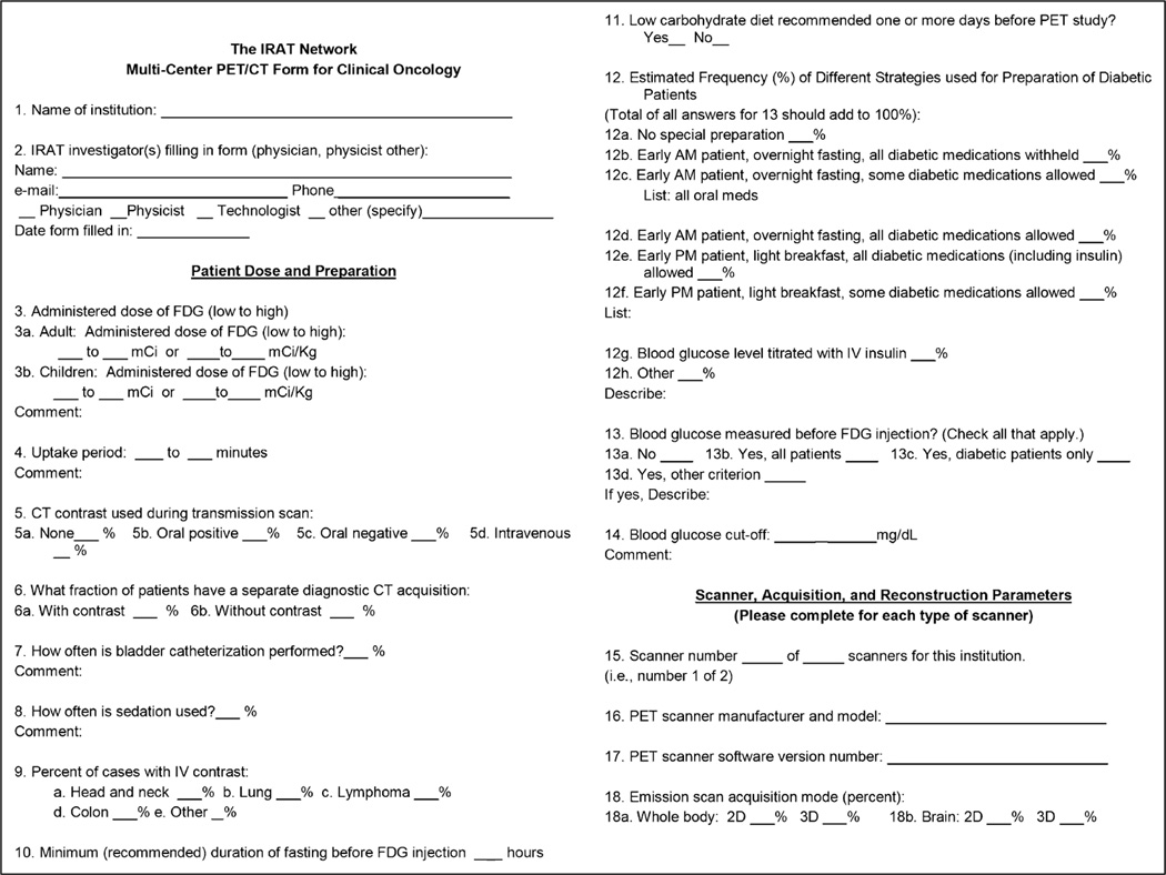
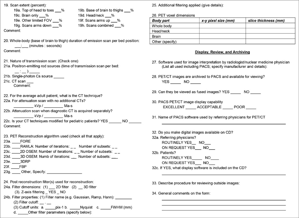
Methods: A 34-question instrument evaluating patient preparation, scanner type, performance approach, display, and analysis was developed. Fifteen institutions, including the 8 original IRATs and 7 institutions that had developed affiliate IRATs, were surveyed.
Results: The major areas of variation were (18)F-FDG dose (259-740 MBq [7-20 mCi]) uptake time (45-90 min), sedation (never to frequently), handling of diabetic patients, imaging time (2-7 min/bed position), performance of diagnostic CT scans as a part of PET/CT, type of acquisition (2-dimensional vs. 3-dimensional), CT technique, duration of fasting (4 or 6 h), and (varying widely) acquisition, processing, display, and PACS software--with 4 sites stating that poor-quality images appear on PACS.
Conclusion: There is considerable variability in the way PET/CT scans are performed at academic institutions that are part of the IRAT network. This variability likely makes it difficult to quantitatively compare studies performed at different centers. These data suggest that additional standardization in methodology will be required so that PET/CT studies, especially those performed quantitatively, are more comparable across sites.
Figures
Comment in
-
Standardization of quantitative imaging: the time is right, and 18F-FDG PET/CT is a good place to start.J Nucl Med. 2011 Feb;52(2):171-2. doi: 10.2967/jnumed.110.081224. Epub 2011 Jan 13. J Nucl Med. 2011. PMID: 21233179 No abstract available.
References
-
- Maziak DE, Darling GE, Inculet RI, et al. Positron emission tomography in staging early lung cancer: a randomized trial. Ann Intern Med. 2009;151:221–228. - PubMed
-
- Fischer B, Lassen U, Mortensen J, et al. Preoperative staging of lung cancer with combined PET-CT. N Engl J Med. 2009;361:32–39. - PubMed
-
- van Tinteren H, Hoekstra OS, Smit EF, et al. Effectiveness of positron emission tomography in the preoperative assessment of patients with suspected non-small-cell lung cancer: the PLUS multicentre randomised trial. Lancet. 2002;359:1388–1393. - PubMed
-
- Shankar LK, Hoffman JM, Bacharach S, et al. Consensus recommendations for the use of 18F-FDG PET as an indicator of therapeutic response in patients in National Cancer Institute Trials. J Nucl Med. 2006;47:1059–1066. - PubMed
-
- Boellaard R, Oyen WJ, Hoekstra CJ, et al. The Netherlands protocol for standardisation and quantification of FDG whole body PET studies in multi-centre trials. Eur J Nucl Med Mol Imaging. 2008;35:2320–2333. - PubMed
Publication types
MeSH terms
Substances
Grants and funding
- P30 CA093373/CA/NCI NIH HHS/United States
- P30 CA023074/CA/NCI NIH HHS/United States
- P30 CA006973/CA/NCI NIH HHS/United States
- P30 CA047904/CA/NCI NIH HHS/United States
- 3P30CA093373/CA/NCI NIH HHS/United States
- P30 CA086862/CA/NCI NIH HHS/United States
- 5P30CA086862/CA/NCI NIH HHS/United States
- 3P30CA047904/CA/NCI NIH HHS/United States
- P30 CA008748/CA/NCI NIH HHS/United States
- 5P30CA091842/CA/NCI NIH HHS/United States
- P30 CA091842/CA/NCI NIH HHS/United States
- P30 CA016058/CA/NCI NIH HHS/United States
- 3P30CA008748/CA/NCI NIH HHS/United States
- 3P30CA016058/CA/NCI NIH HHS/United States
- 2P30CA023074/CA/NCI NIH HHS/United States
- 3P30CA006973/CA/NCI NIH HHS/United States
LinkOut - more resources
Full Text Sources
Other Literature Sources