. 2011 Mar 25;286(12):10690-701.
doi: 10.1074/jbc.M110.202507. Epub 2011 Jan 13.

Crystal structures of poly(ADP-ribose) polymerase-1 (PARP-1) zinc fingers bound to DNA: structural and functional insights into DNA-dependent PARP-1 activity

Affiliations

Crystal structures of poly(ADP-ribose) polymerase-1 (PARP-1) zinc fingers bound to DNA: structural and functional insights into DNA-dependent PARP-1 activity

Marie-France Langelier et al. J Biol Chem. .

Abstract

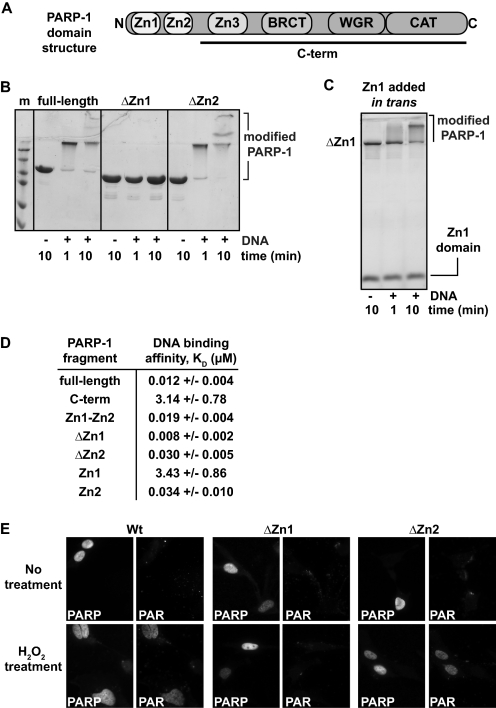
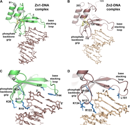
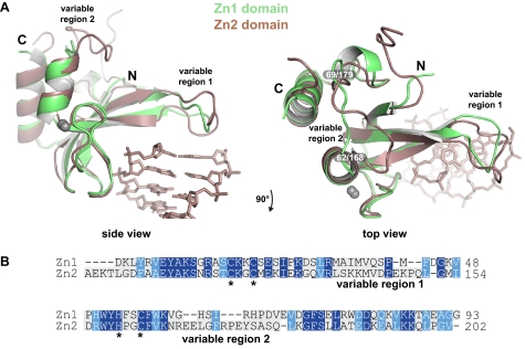
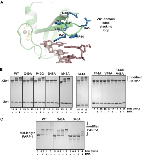
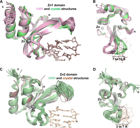
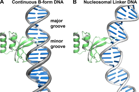
Poly(ADP-ribose) polymerase-1 (PARP-1) has two homologous zinc finger domains, Zn1 and Zn2, that bind to a variety of DNA structures to stimulate poly(ADP-ribose) synthesis activity and to mediate PARP-1 interaction with chromatin. The structural basis for interaction with DNA is unknown, which limits our understanding of PARP-1 regulation and involvement in DNA repair and transcription. Here, we have determined crystal structures for the individual Zn1 and Zn2 domains in complex with a DNA double strand break, providing the first views of PARP-1 zinc fingers bound to DNA. The Zn1-DNA and Zn2-DNA structures establish a novel, bipartite mode of sequence-independent DNA interaction that engages a continuous region of the phosphodiester backbone and the hydrophobic faces of exposed nucleotide bases. Biochemical and cell biological analysis indicate that the Zn1 and Zn2 domains perform distinct functions. The Zn2 domain exhibits high binding affinity to DNA compared with the Zn1 domain. However, the Zn1 domain is essential for DNA-dependent PARP-1 activity in vitro and in vivo, whereas the Zn2 domain is not strictly required. Structural differences between the Zn1-DNA and Zn2-DNA complexes, combined with mutational and structural analysis, indicate that a specialized region of the Zn1 domain is re-configured through the hydrophobic interaction with exposed nucleotide bases to initiate PARP-1 activation.


Figures

FIGURE 1.
FIGURE 1.
The Zn1 and Zn2 domains of PARP-1 have distinct biochemical activities. A, schematic representation of PARP-1 domain structure. B, DNA-dependent automodification assay of full-length PARP-1, ΔZn1, and ΔZn2 (1 μm) in the presence of 1 μm DNA and 5 mm NAD+. Time points were analyzed by 12% SDS-PAGE. C, DNA-dependent automodification assay of ΔZn1 PARP-1 in the presence/absence of the isolated Zn1 domain added in trans. Time points were analyzed by 15% SDS-PAGE. D, DNA binding affinity constants (KD) derived from fluorescence polarization experiments using an 18-bp DNA. The values represent the average and mean ± S.D. of three or more independent experiments. E, PARP-1−/− mouse embryonic fibroblasts were transiently transfected with an expression vector coding for PARP-1 full-length, ΔZn1 or ΔZn2. Fixed cells were stained with antibodies against PAR or PARP-1 as indicated. Top panels, no treatment; bottom panels, treatment with H2O2 for 10 min.
FIGURE 2.
FIGURE 2.
Crystal structures of PARP-1 zinc fingers in complex with DNA demonstrate the molecular basis for structure-specific DNA binding. A, x-ray structure of the Zn1 domain bound to a 10-bp DNA duplex (DNA sequence G is shown). For clarity, the Zn1 molecule bound to the other end of the DNA is not shown (see supplemental Fig. S3). Zinc-coordinating residues are drawn as yellow sticks; the zinc atom is drawn as a gray sphere. B, x-ray structure of the Zn2 domain bound to an 8-bp DNA duplex (DNA sequence B is shown). Zinc-coordinating residues are drawn as yellow sticks; the zinc atom is drawn as a gray sphere. C and D, a more detailed view of the backbone grip and base stacking loop of the Zn1 and Zn2 domains, respectively. Residues mentioned in the text are drawn as blue sticks.
FIGURE 3.
FIGURE 3.
Alignment of the Zn1 and Zn2 domain structures in complex with DNA. A, two views of an alignment of the Zn1 and Zn2 domains, highlighting two structurally distinct regions, labeled variable region 1 (base stacking loop) and variable region 2. Only the DNA duplex from the Zn1-DNA structure is shown for clarity. B, structure-based amino acid sequence alignment of human PARP-1 Zn1 and Zn2 domains. Conserved residues are shaded blue; zinc-coordinating residues are marked with stars.
FIGURE 4.
FIGURE 4.
Specific residues of the Zn1 base stacking loop are required for DNA-dependent PARP-1 activation. A, detailed view of the Zn1 base stacking loop. B, DNA-dependent automodification assay using a combination of WT or mutant Zn1 domain, the ΔZn1 fragment of PARP-1, 5 mm NAD+, and the absence or presence of 1 μm DNA. Time points were analyzed on 15% SDS-PAGE with Coomassie staining. C, DNA-dependent automodification activity of full-length PARP-1 bearing mutations in the Zn1 domain. Time points were analyzed on 12% SDS-PAGE with Coomassie staining.
FIGURE 5.
FIGURE 5.
The zinc finger base stacking loop is repositioned upon binding to DNA. A, the 20 deposited NMR models of the Zn1 domain in the absence of DNA (PDB code 2dmj; pink) aligned with the 8 Zn1 domains present in the Zn1-DNA complex x-ray structure (green). B, down view of the alignment in panel A. C, the 20 deposited NMR models of the Zn2 domain in the absence of DNA (PDB 2cs2; pale green) aligned with the 2 Zn2 domains in the Zn2-DNA complex x-ray structure (brown). D, down view of the alignment in panel C.
FIGURE 6.
FIGURE 6.
The zinc finger base stacking loop inserts into the major groove of continuous DNA structures. A, continuous B-form DNA (light gray) aligned to the DNA duplex in the Zn1-DNA crystal structure (dark gray) highlights that the base stacking loop inserts into the DNA major groove. DNA distortions will be required to fully accommodate the base stacking loop. B, the Zn1 domain modeled on a segment of bent nucleosomal linker DNA illustrates the type of DNA distortion that is anticipated to interact with PARP-1 zinc fingers. DNA bending toward the minor groove better accommodates the base stacking loop compared with B-form DNA (DNA extracted from PDB coordinate 1zbb (45)).

References

    1. Hassa P. O., Hottiger M. O. (2008) Front. Biosci. 13, 3046–3082 - PubMed
    1. Krishnakumar R., Kraus W. L. (2010) Mol. Cell 39, 8–24 - PMC - PubMed
    1. D'Amours D., Desnoyers S., D'Silva I., Poirier G. G. (1999) Biochem. J. 342, 249–268 - PMC - PubMed
    1. Rouleau M., Patel A., Hendzel M. J., Kaufmann S. H., Poirier G. G. (2010) Nat. Rev. Cancer 10, 293–301 - PMC - PubMed
    1. Haince J. F., McDonald D., Rodrigue A., Déry U., Masson J. Y., Hendzel M. J., Poirier G. G. (2008) J. Biol. Chem. 283, 1197–1208 - PubMed

Publication types