. 2011 Feb 1;108(5):2010-5.
doi: 10.1073/pnas.1018974108. Epub 2011 Jan 13.

CD8+ T regulatory cells express the Ly49 Class I MHC receptor and are defective in autoimmune prone B6-Yaa mice

Affiliations

CD8+ T regulatory cells express the Ly49 Class I MHC receptor and are defective in autoimmune prone B6-Yaa mice

Hye-Jung Kim et al. Proc Natl Acad Sci U S A. .

Abstract

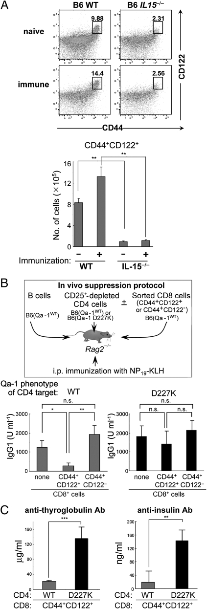
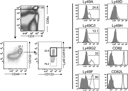
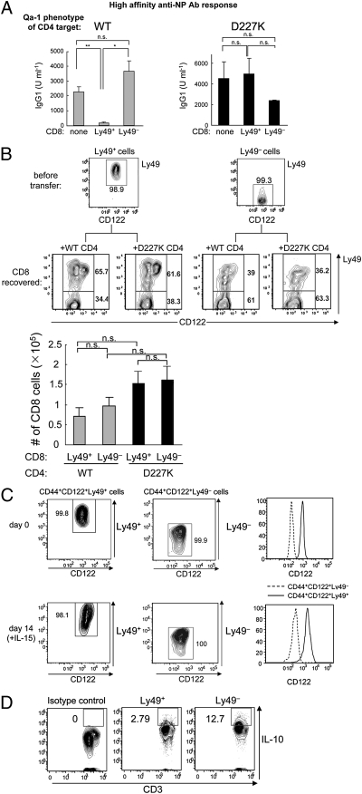
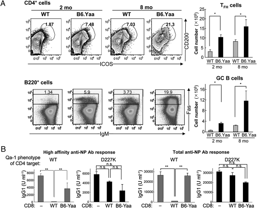
The immune system includes a subpopulation of CD8(+) T cells equipped to inhibit the expansion of follicular T helper (T(FH)) cells, resulting in suppression of autoantibody production and associated lupus-like disease. These CD8(+) T regulatory (Treg) cells recognize Qa-1/peptide complexes on target T(FH) cells and depend on the IL-15 cytokine for development and function. Here we show that these CD8(+) Treg cells express a triad of surface receptors--CD44, CD122, and the class I MHC receptor Ly49--and account for <5% of CD8(+) T cells. Moreover, the development of systemic lupus erythematosus-like disease in B6-Yaa mutant mice is associated with a pronounced defect in CD8(+) Treg cell activity, suggesting that this regulatory subset may represent an effective therapeutic approach to systemic lupus erythematosus-like autoimmune disease.


Conflict of interest statement

The authors declare no conflict of interest.

Figures

Fig. 1.
Fig. 1.
Qa-1–dependent suppression of Ab response by CD44+CD122+ CD8 T cells. (A) The percentage and number of CD44+CD122+ CD8 T cells from naïve and KLH/CFA-immunized (day 8) WT or IL-15−/− mice. FACS profiles after gating on CD3+CD8+ cells are shown. (B) Ab response after transfer of B, CD4, and CD8 cells into Rag2−/− mice. Here 2 × 10⁶ WT naïve B cells were transferred along with 1 × 10⁶ CD25+ depleted CD4 cells from B6.Qa-1(WT) or B6.Qa-1(D227K) mice into Rag2−/− hosts. CD44+CD122+ or CD44+CD122 CD8+ T cells isolated from KLH/CFA immunized WT B6 mice were also transferred into these Rag2−/− hosts before recipients were immunized i.p. with 100 μg of NP19-KLH in CFA. At day 10, mice were challenged i.p. with 50 μg of NP19-KLH in IFA, and NP-specific Ab responses were measured by ELISA 7 d later. (C) Defective inhibition by CD8 Treg cells leads to autoantibody generation. Anti-thyroglobulin and anti-insulin Abs generated in Rag2−/− recipients were measured by ELISA at day 50. Error bars denote mean ± SEM.
Fig. 2.
Fig. 2.
Ly49 expression by CD8+ Treg cells after immunization. WT B6 mice were immunized i.p. with 100 μg of KLH in CFA, and 10 d later the surface expression of CD44, CD122, Ly49 subtypes, CD8β, and CD62L was analyzed. Staining of Ly49+ cells was done using the following Abs: Ly49C/I/F/H (clone 4B11), Ly49C/I (clone 5E6), Ly49A (clone A1), and Ly49G2 (clone eBio4D11). The numbers represent the percentage of cells expressing each surface protein.
Fig. 3.
Fig. 3.
CD44+CD122+Ly49+ CD8 T cells account for Qa-1–restricted suppressive activity. (A) Ab response after transfer of B, CD4, and CD8 cells into Rag2−/− hosts. Here 2 × 10⁶ WT naïve B cells were transferred along with 0.5 × 10⁶ CD25+-depleted CD4 cells from B6.Qa-1(WT) or B6.Qa-1(D227K) mice into Rag2−/− hosts. CD44+CD122+Ly49+ CD8+ T cells were isolated from KLH/CFA-immunized WT B6 mice, and 0.15 × 10⁶ cells were transferred into Rag2−/− hosts. Immediately after cell transfer, the Rag2−/− recipients were immunized i.p. with 100 μg of NP19-KLH in CFA. At day 10, mice were challenged i.p. with 50 μg of NP19-KLH in IFA, and high-affinity NP-specific Ab responses were measured by ELISA 7 d after the challenge. Data shown represent mean ± SEM. (B) Phenotype of CD8 T cells in Rag2−/− hosts transferred with either CD44+CD122+Ly49+ or CD44+CD122+Ly49 CD8+ T cells along with WT B cells and CD4 T cells (WT or D227K) were analyzed for surface Ly49 expression 3 wk after cell transfer. (Upper) Numbers shown in the gate represent the percentage of cells. (Lower) Total CD8 T cell numbers recovered from Rag2−/− hosts reconstituted with B, CD4, and CD8 cells from different origins are shown. (C) Phenotypic stability of Ly49+ and Ly49 CD8 T cells in vitro. CD44+CD122+Ly49+ and CD44+CD122+Ly49 CD8 T cells were sorted by FACS (purity >99%). Cells were incubated with 100 ng/mL of IL-15 for 14 d, after which the phenotype was reanalyzed. The percentages of Ly49+ and Ly49 cells and levels of CD122 expression in each cell subset are shown. (D) Expression of IL-10 by Ly49+ and Ly49 CD8 cells. Ly49+ and Ly49 CD8 cells were stimulated with phorbol myristate acetate/ionomycin for 8 h, and the production of IL-10 was measured by intracellular cytokine staining. The percentages of IL-10+ Ly49+ and Ly49 cells are shown. Immunofluorescence of Ly49 cells labeled with isotype control Ab is shown as a negative control.
Fig. 4.
Fig. 4.
Expansion of TFH and GC B cells in B6-Yaa mice. (A) Spleen cells from age-matched (2 mo and 8 mo) WT B6 and B6-Yaa mice were stained with CD4, ICOS, and CD200 antibodies for TFH cells and with B220, IgM, and Fas antibodies for GC B cells. (Right) The absolute numbers of TFH and GC B cells are shown. (B) Impaired suppression of Ab response by CD8+ T cells from B6-Yaa mice. WT naïve B cells were transferred along with CD25+-depleted CD4 T cells from Qa-1 (WT) or Qa-1 (D227K) mice into Rag2−/− hosts. FACS-sorted CD44+CD122+ CD8 T cells isolated from KLH/CFA-immunized WT or B6-Yaa mice were transferred into Rag2−/− hosts. Immediately after cell transfer, the Rag2−/− recipients were immunized i.p. with 100 μg of NP19-KLH in CFA and then reimmunized i.p. with 50 μg of NP19-KLH in IFA 20 d later. Secondary Ab responses for high-affinity and total anti-NP IgG1 are shown.

References

    1. Bouneaud C, Kourilsky P, Bousso P. Impact of negative selection on the T cell repertoire reactive to a self-peptide: A large fraction of T cell clones escapes clonal deletion. Immunity. 2000;13:829–840. - PubMed
    1. Goldrath AW, Bevan MJ. Selecting and maintaining a diverse T-cell repertoire. Nature. 1999;402:255–262. - PubMed
    1. Slifka MK, et al. Preferential escape of subdominant CD8+ T cells during negative selection results in an altered antiviral T cell hierarchy. J Immunol. 2003;170:1231–1239. - PubMed
    1. Martin DA, et al. Defective CD95/APO-1/Fas signal complex formation in the human autoimmune lymphoproliferative syndrome, type Ia. Proc Natl Acad Sci USA. 1999;96:4552–4557. - PMC - PubMed
    1. Kearney ER, Pape KA, Loh DY, Jenkins MK. Visualization of peptide-specific T cell immunity and peripheral tolerance induction in vivo. Immunity. 1994;1:327–339. - PubMed

Publication types

MeSH terms

Substances