. 2011 Feb 1;108(5):2058-63.
doi: 10.1073/pnas.1018157108. Epub 2011 Jan 13.

High-throughput ectopic expression screen for tamoxifen resistance identifies an atypical kinase that blocks autophagy

Affiliations

High-throughput ectopic expression screen for tamoxifen resistance identifies an atypical kinase that blocks autophagy

Laura Gonzalez-Malerva et al. Proc Natl Acad Sci U S A. .

Abstract

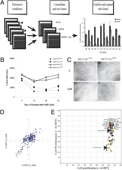
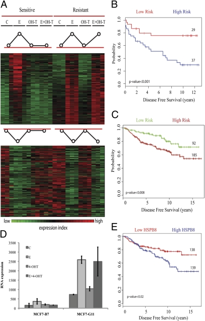
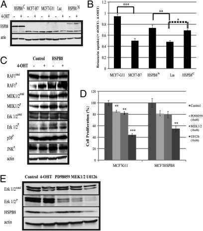
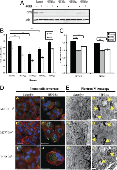
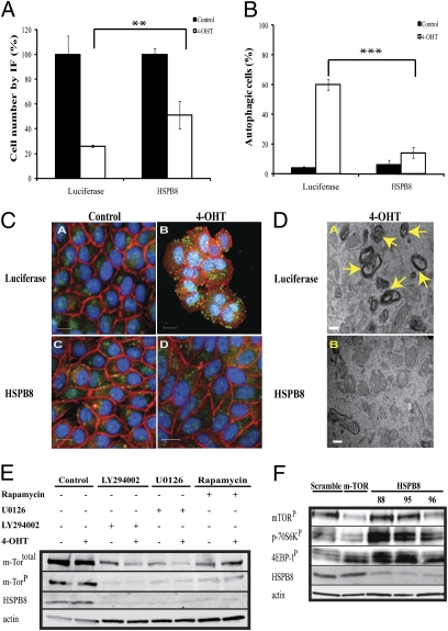
Resistance to tamoxifen in breast cancer patients is a serious therapeutic problem and major efforts are underway to understand underlying mechanisms. Resistance can be either intrinsic or acquired. We derived a series of subcloned MCF7 cell lines that were either highly sensitive or naturally resistant to tamoxifen and studied the factors that lead to drug resistance. Gene-expression studies revealed a signature of 67 genes that differentially respond to tamoxifen in sensitive vs. resistant subclones, which also predicts disease-free survival in tamoxifen-treated patients. High-throughput cell-based screens, in which >500 human kinases were independently ectopically expressed, identified 31 kinases that conferred drug resistance on sensitive cells. One of these, HSPB8, was also in the expression signature and, by itself, predicted poor clinical outcome in one cohort of patients. Further studies revealed that HSPB8 protected MCF7 cells from tamoxifen and blocked autophagy. Moreover, silencing HSBP8 induced autophagy and caused cell death. Tamoxifen itself induced autophagy in sensitive cells but not in resistant ones, and tamoxifen-resistant cells were sensitive to the induction of autophagy by other drugs. These results may point to an important role for autophagy in the sensitivity to tamoxifen.


Conflict of interest statement

The authors declare no conflict of interest.

Figures

Fig. 1.
Fig. 1.
MCF7 subclones and kinome screen. (A) MCF7 cells were diluted and plated into 96-well plates. Condensed plates were split into three different plates: archive, control, and tamoxifen. Cell proliferation was measured and 12 subclones were selected. (B) Cell proliferation assay showed that resistant subclones continued to proliferate in presence of 4-OHT (1 μM) and tamoxifen-sensitive cells were inhibited. (C) Photomicrographs of MCF7-B7TamS and MCF7-G11TamR showed growth inhibition of sensitive but not resistant cells. (D) Scatter plot of the z-scores showing the correlation of results for kinase cDNA expression on cell growth of MCF7-C11TamS after 4-OHT at 1 and 5 μM and puromycin (1 μg/mL). (E) Scatter plot of cell proliferation based on resazurin of 80 candidate kinases in the presence and absence of 4-OHT (1 μM) and puromycin.
Fig. 2.
Fig. 2.
Microarray expression profiles of tamoxifen-sensitive and -resistant subclones. (A) Gene cluster analysis for E-regulated genes showed 115 induced and 112 repressed genes, which no longer respond to tamoxifen in resistant cells. Stimulated genes are shown in red, inhibited genes in green, with black intermediate between the two. (B) From the genes in A, a set of 67 genes were selected to test prognostic significance. Kaplan-Meier survival analysis from Miller's dataset (18) showed 37 patients with early recurrence compared with 29 showing better disease-free survival; and (C) 92 patients showed better disease-free survival compared with 185 at high risk in Loi's dataset (19). (D) Expression of HSPB8 increased in both cell lines after E addition, but is suppressed by tamoxifen in MCF7-B7TamS and not in MCF7-G11TamR. (E) A Kaplan-Meier curve for disease-free survival, sorted based only on HSPB8 expression, showed better outcome when expression is low. From this set, 67 of the 92 low-risk patients showed low expression of HSPB8 (73% overlap). Similarly, 114 of the 139 patients with high HSPB8 expression were in the high-risk group (82% overlap).
Fig. 3.
Fig. 3.
Stable ectopic expression of HSPB8 blocks autophagy after tamoxifen. (A) Stable cell lines in MCF7-B7TamS expressing HSPB8 flag-tagged at either HSPB8C or HSPB8N were compared by immunoblot with MCF7-B7TamS and MCF7-G11TamR after 4-OHT (1 μM) for 72 h. Luciferase (Luc) was used as a negative control. (B) Cell proliferation assay of above cells was performed in 96-well plates. Data represent resazurin uptake in presence of 4-OHT divided by resazurin uptake in absence of drug. (*P < 0.07, **P < 0.05, ***P < 0.005 calculated based on Welch Two Samples t test.) (C) Western blot analysis of the phosphorylation status of MAP kinases. RAFP and ERK1/2P were elevated in HSPB8 and increased further in presence of 4-OHT. (D) Stable HSPB8 and MCF7-G11TamR cells were treated for 72 h with three MEK inhibitors: PD98059, MEK1/2, and U0126 and control (**P < 0.01, ***P < 0.00001). (E) ERK1/2P and HSPB8 were measured by western-blot after MCF7-G11TamR was treated with MEK inhibitors.
Fig. 4.
Fig. 4.
Reducing HSPB8 in MCF7-G11TamR leads to autophagy. (A) Protein expression of HSPB8 was reduced with three of four different targeted hairpins in MCF7-G11TamR compared with vector-expressing scrambled sequences. (B) Silencing HSPB8 reduced cell proliferation of MCF7-G11TamR. The effect was more pronounced in the presence of 4-OHT (1 μM). Resazurin data were normalized against scramble in the presence of puromycin and in absence of 4-OHT after 48 h postdrug treatment (**P < 0.001). (C) Hairpins HSPB888 and HSPB895 reduced cell proliferation compared with scramble in two additional tamoxifen-resistant subclones, MCF7-H9TamR and T47D-G9TamR, in the presence of 4-OHT (**P < 0.001). (D) Immunofluorescence images after silencing HSPB888 in all resistant subclones showed the characteristic redistribution of anti-MAP LC3 into punctuate green structures (B, F, and J, respectively), indicating active autophagy, but scrambled hairpin showed only diffuse staining (A, E, and I). Blue: nucleus by Hoechst; red: actin by Phalloidin; green: autophagy marker anti-MAP LC3. (Scale bars 50 μM.) (E) EM consistently identified a large number of autophagosomes containing organelles undergoing degenerative changes (arrows) in all of the cells where HSPB8 was silenced (D, H, and L). (Scale bars, 500 nm.)
Fig. 5.
Fig. 5.
Effect of autophagy in sensitive and resistant cells. (A) Fluorescence images of quadruplicate experiments were taken after LC3 staining in MCF7-B7TamS cells stably expressing either HSPB8 or luciferase (control). Total number of cells were counted in the presence and absence of 4-OHT (1 μM) after 72 h and expressed as a percentage of no treatment controls (**P < 0.01). (B) HSPB8 reduced the percentage of autophagic cells (mean ± SD) (***P < 0.003). (C) Representative images from B showing reduction of autophagy. Blue: nucleus by Hoechst; red: actin by Phalloidin; green: autophagy marker anti-MAP LC3. (Scale bars, 50 μM.) (D) EM images showed the presence of clusters of autophagosomes after control cells were treated with 4-OHT. Autophagosomes were markedly reduced when the cells were ectopically expressing HSPB8. (Scale bar, 500 nm.) (E) Western blot analysis showed lower expression levels of mTOR and HSPB8 in MCF7-G11TamR 72 h posttreatment with autophagy-inducing drugs: rapamycin (0.1 μM), LY294002 (5 μM), U0126 (10 μM), but not with 4-OHT (1 μM). (F) Short hairpin RNAs directed at HSPB8, mTOR, or scramble were transduced into MCF7-G11TamR cells. Protein expression levels of HSPB8, mTORP, p70S6kP, and 4EBP-1P were measured 72 h postinfection. β-Actin served as a loading control.

References

    1. Musgrove EA, Sutherland RL. Biological determinants of endocrine resistance in breast cancer. Nat Rev Cancer. 2009;9:631–643. - PubMed
    1. Riggins RB, Schrecengost RS, Guerrero MS, Bouton AH. Pathways to tamoxifen resistance. Cancer Lett. 2007;256(1):1–24. - PMC - PubMed
    1. Ring A, Dowsett M. Mechanisms of tamoxifen resistance. Endocr Relat Cancer. 2004;11:643–658. - PubMed
    1. Yu J, et al. A transcriptional fingerprint of estrogen in human breast cancer predicts patient survival. Neoplasia. 2008;10(1):79–88. - PMC - PubMed
    1. Frasor J, et al. Gene expression preferentially regulated by tamoxifen in breast cancer cells and correlations with clinical outcome. Cancer Res. 2006;66:7334–7340. - PubMed

Publication types

MeSH terms