. 2011 Jun;18(6):1046-56.
doi: 10.1038/cdd.2010.177. Epub 2011 Jan 14.

Mortalin-p53 interaction in cancer cells is stress dependent and constitutes a selective target for cancer therapy

Affiliations

Mortalin-p53 interaction in cancer cells is stress dependent and constitutes a selective target for cancer therapy

W-J Lu et al. Cell Death Differ. 2011 Jun.

Abstract

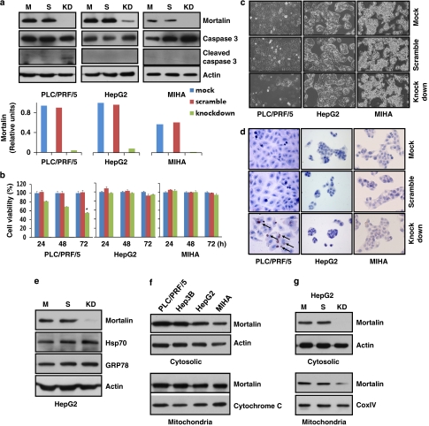
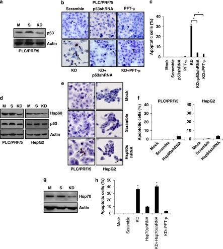
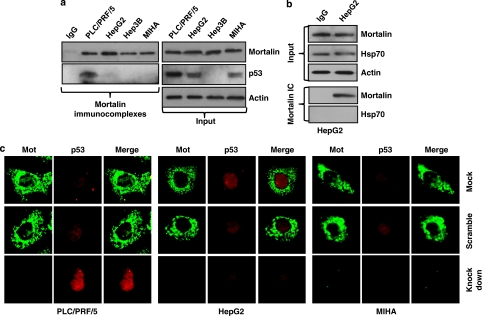
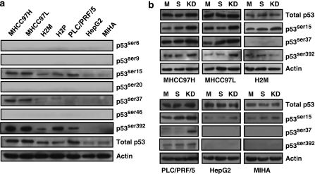
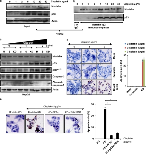
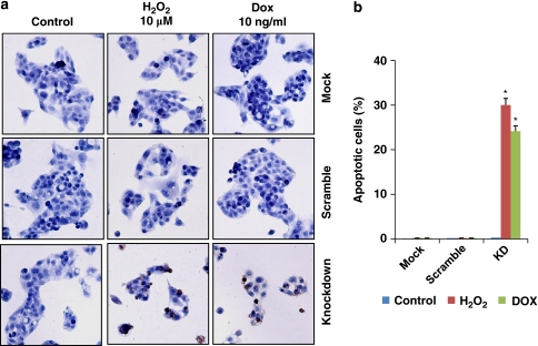
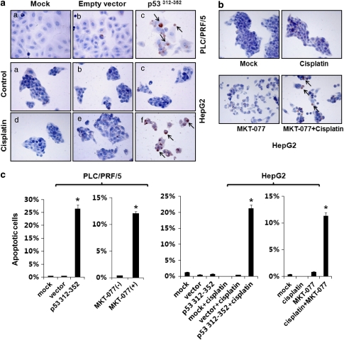
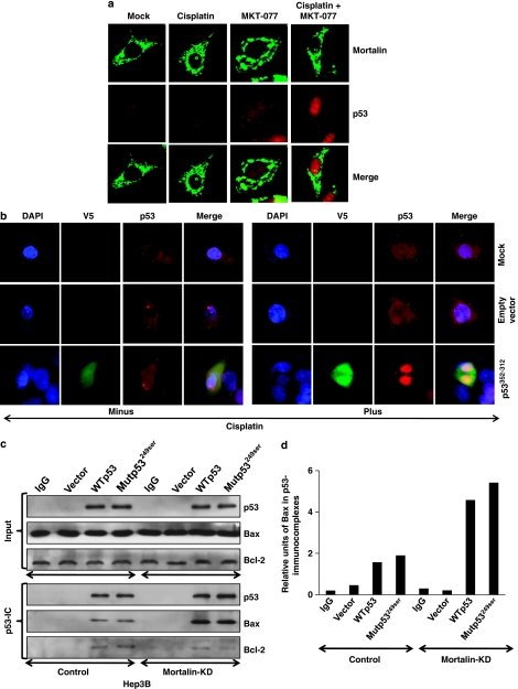
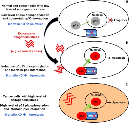
Stress protein mortalin is a multifunctional protein and is highly expressed in cancers. It has been shown to interact with tumor suppressor protein-p53 (both wild and mutant types) and inactivates its transcriptional activation and apoptotic functions in cancer cells. In the present study, we found that, unlike most of the cancer cells, HepG2 hepatoma lacked mortalin-p53 interaction. We demonstrate that the mortalin-p53 interaction exists in cancer cells that are either physiologically stressed (frequently associated with p53 mutations) or treated with stress-inducing chemicals. Targeting mortalin-p53 interaction with either mortalin small hairpin RNA or a chemical or peptide inhibitor could induce p53-mediated tumor cell-specific apoptosis in hepatocellular carcinoma; p53-null hepatoma or normal hepatocytes remain unaffected.


Figures

Figure 1
Figure 1
Mortalin silencing induces apoptosis in PLC/PRF/5, but not in MIHA and HepG2 cells. (a) Mortalin shRNA caused reduction in mortalin expression (KD) in all the three cell lines (examined at 72 h post transfection); empty vector transfected (Mock-M) and scrambled shRNA transfected (S) cells were used as control. (b) Viability (MTT assay) was significantly reduced in PLC/PRF/5 (*P<0.05) at 72 h post-transfection of mortalin shRNA; MIHA cells and HepG2 did not show any change as compared with controls. (c) Phase-contrast images of control and mortalin shRNA transfected cells showing apoptosis in mortalin shRNA transfected PLC/PRF/5 cells only. (d) In situ TUNEL staining identified mortalin shRNA-induced apoptosis in PLC/PRF/5, but not in MIHA and HepG2 cells. Apoptotic PLC cells are marked with arrows. (e) Mortalin knockdown cells (KD) were examined for Hsp70 and glucose-regulated protein 78 (GRP78) by western blotting with specific antibodies. The mortalin shRNA-2166 was highly specific for mortalin and did not cause any reduction in Hsp70 and GRP78. (f) Presence of mortalin in the cytoplasmic and mitochondrial fractions in four cell lines was examined by western blotting with mortalin specific antibody. (g) Mortalin knockdown by shRNA-2166 caused reduction in mortalin in both cytosolic and mitochondrial fractions
Figure 2
Figure 2
Mortalin silencing-induced apoptosis could be rescued by p53 shRNA and p53-specific inhibitor. (a) Western blot showing p53 knockdown with p53 shRNA in PLC/PRF/5 cells. (b) In situ TUNEL staining showing that mortalin knockdown (KD)-induced apoptosis was significantly compromised by treatment with p53 shRNA and p53-specific inhibitor (PFT-μ) in PLC/PRF/5 cells. (c) Quantitation of apoptotic cells is shown (*P<0.05). (d) Western blot showing Hsp60 knockdown. (e) In situ TUNEL staining showing that Hsp60 knockdown did not induce apoptosis. (f) Quantitation of apoptotic cells is shown. (g) Cells transfected with Hsp70 shRNA showed reduction in Hsp70 protein. (h) Mortalin knockdown (KD) showed higher level of apoptosis as compared with the Hsp70 knockdown cells, and mortalin knockdown-induced apoptosis was recovered by co-treatment with PFT-μ cells (*P<0.05). Arrows in b and e show the apoptotic cells
Figure 3
Figure 3
Mortalin–p53 interaction was detected only in PLC, but not in MIHA and HepG2 cells. (a) p53 was detected in mortalin immunocomplexes in PLC/PRF/5. HepG2, Hep3B and MIHA cells lacked this interaction. (b) Mortalin antibody used for immunoprecipitation was specific for mortalin and did not crossreact with Hsp70. (c) Mortalin (green) knockdown induced nuclear translocation of p53 (red) in PLC cells. HepG2 and MIHA cells did not show nuclear translocation of p53 after mortalin knockdown (examined at 72 h post-transfection)
Figure 4
Figure 4
Mortalin shRNA-induced apoptosis depends on mortalin–p53 interaction that in turn depends on the level of cellular stress reflected by p53 phosphorylation. (a) p53 phosphorylation at seven common phosphorylation sites was detected in six HCC and one normal immortalized liver cell line, MIHA. Strong phosphorylation signals at three phosphorylation sites (Ser 15, Ser 37, Ser 392) were detected in 97H, 97L, H2M, H2P and PLC/PRF/5 as compared with HepG2 and MIHA cells, suggesting that the former four cell lines represent cases of higher physiological stress level. (b) Mortalin shRNA-2166 caused an increase in p53 phosphorylation at one or more sites in four HCC cell lines; HepG2 and MIHA did not show any change in p53 phosphorylation (examined at 72 h post-transfection)
Figure 5
Figure 5
Mortalin–p53 interaction is induced by stress. (a) Exposure of HepG2 cells to cisplatin caused increase in p53 and mortalin. (b) Mortalin–p53 interaction was induced in HepG2 cells by cisplatin treatment (48 h). (c) HepG2 cells were sensitized to mortalin shRNA-induced apoptosis after cisplatin stress-induced mortalin–p53 interactions. Cisplatin-treated cells showed increase in p53-serine15 phosphorylation and caspase 3 cleavage when co-treated with mortalin shRNA-2166 (KD). (d) In situ TUNEL staining of HepG2 cells corresponding to Figure 3c. Apoptosis was observed in cisplatin (1 and 2 μg/ml) and mortalin shRNA-2166 (knockdown) cells only. Quantitation of apoptotic cells is shown (*P<0.05). (e) Apoptosis induced by mortalin knockdown (KD) and cisplatin treatment was rescued by either p53 knockdown with p53 shRNA or with a specific inhibitor of p53 (PFT-μ)
Figure 6
Figure 6
HepG2 cells were sensitized to mortalin knockdown-induced apoptosis by stress treatment. (a) In situ TUNEL staining showing both H2O2 (oxidative stress) and doxorubicin (DNA damage stress) treatment (48 h) caused apoptosis of HepG2 cells in combination with mortalin shRNA. (b) Quantitation of apoptotic cells is shown (*P<0.05)
Figure 7
Figure 7
Reactivation of the apoptotic function of p53 by targeting mortalin–p53 interaction with and without stress. Mortalin–p53 interaction was targeted by mortalin–p53 binding antagonists, mortalin-binding peptide p53312−352 and cationic dye MKT-077. (a) PLC/PRF/5 cells underwent apoptosis in response to the expression of p53312−352 peptide. HepG2 cells (lacking mortain–p53 interactions) were sensitized to p53312−352-induced apoptosis by cisplatin treatment. (b) HepG2 cells showed no apoptosis in response to either cisplatin or MKT-077 treatment alone. Apoptosis was induced by co-treatment with cisplatin and MKT-077. (c) Quantitation of apoptotic cells was shown (*P<0.05)
Figure 8
Figure 8
Nuclear accumulation of p53 in HepG2 cells. HepG2 cells treated with either MKT-077 (a) or peptides inhibitor (b) showed nuclear accumulation of p53, only when co-treated with cisplatin. (c) Mortalin knockdown cells (with wild-type or mutant p53) showed increased p53–Bax interaction; the quantitation of Bax present in p53 immunocomplexes in control and mortalin knockdown cells is shown in (d)
Figure 9
Figure 9
A schematic model showing the effect of mortalin knockdown and nuclear translocation of p53 in cells with different levels of stress. (a) In normal, immortalized and non-malignant cancer cells with low p53 phosphorylation, mortalin does not interact with p53, and hence p53 freely translocates from cytoplasm to nucleus. However, unphosphorylated p53 is inactive, unstable and insufficient for apoptotic activity. Mortalin knockdown has no effect in this category of cells. (b) Exposure of cells to stress (genotoxic or malignancy) causes p53 phosphorylation and accumulation that induces mortalin–p53 interaction. Mortalin silencing by shRNA in this scenario causes nuclear translocation of the phosphorylated p53 that is stable and active, resulting in apoptotic death of cells. (c) In malignant cancer cells with high stress, p53 is heavily phosphorylated and stable. In these cells, mortalin captures p53 in the cytoplasm and blocks its transcriptional activation, growth arrest and apoptotic functions. In this scenario, mortalin knockdown causes nuclear translocation of p53, leading to apoptotic death of cells. According to the proposed model, mortalin–p53 interaction is selective for stressed cancer cells, and hence could serve as a safe target for cancer therapeutics

References

    1. Kastan MB, Zhan QM, Eldeiry WS, Carrier F, Jacks T, Walsh WV, et al. A mammalian cell cycle checkpoint pathway utilizing p53 and GADD45 is defective in ataxia-telangiectasia. Cell. 1992;71:587–597. - PubMed
    1. Yonish-Rouach E, Resnitzky D, Lotem J, Sachs L, Kimchi A, Oren M. Wild-type p53 induces apoptosis of myeloid leukaemic cells that is inhibited by interleukin-6. Nature. 1991;352:345–347. - PubMed
    1. Nikolaev AY, Li M, Puskas N, Qin J, Gu W. Parc: a cytoplasmic anchor for p53. Cell. 2003;112:29–40. - PubMed
    1. Zhao LY, Liao DQ. Sequestration of p53 in the cytoplasm by adenovirus type 12 E1B 55-kilodalton oncoprotein is required for inhibition of p53-mediated apoptosis. J Virol. 2003;77:13171–13181. - PMC - PubMed
    1. Liu S, Li J, Tao Y, Xiao X. Small heat shock protein alphaB-crystallin binds to p53 to sequester its translocation to mitochondria during hydrogen peroxide-induced apoptosis. Biochem Biophys Res Commun. 2007;354:109–114. - PubMed

Publication types

MeSH terms