Case Reports
doi: 10.4103/0974-2069.74065.
Twin heart with a fused atria and separate ventricles in conjoined twins
Affiliations
- PMID: 21234207
- PMCID: PMC3017932
- DOI: 10.4103/0974-2069.74065
Item in Clipboard
Case Reports
Twin heart with a fused atria and separate ventricles in conjoined twins
Ann Pediatr Cardiol.
2010 Jul.
Abstract
One of the most interesting congenital malformations is that of conjoined twins. We report echocardiographic features of twin heart in dicephalus, tribrachius, dispinous, thoracoomphalopagus twin. It showed two hearts fused at atrial level. Right-sided heart had single atrial chamber with a single ventricle. A single great vessel, aorta, originated from it. Left-sided heart was well developed with two atria and two ventricles. There was a small mid muscular ventricular septal defect and a small patent ductus arteriosus. Great arteries had normal origins.
Keywords: Cardiac malformations in twins; conjoined twins; fused heart.
Conflict of interest statement
Figures

Conjoined twin
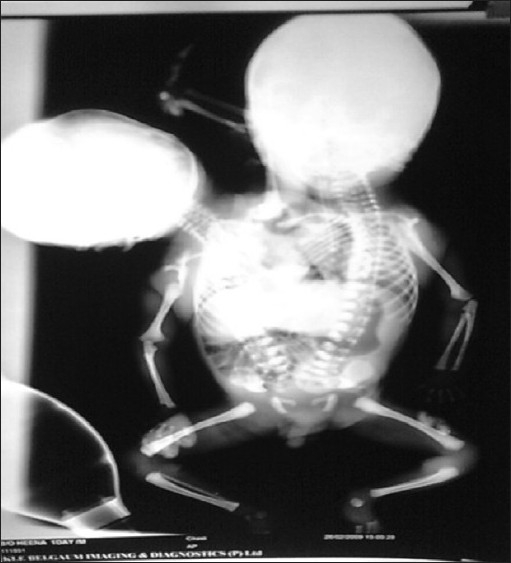
Infantogram showing two heads, two thoraces, two hearts and two vertebral columns, three upper limbs and two lower limbs

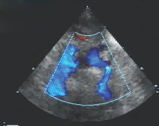
Echo showing two hearts fused at atrial level. Right-sided heart had single atrial chamber communicating with a single ventricle through single atrioventricular valve and left-side heart communicated with two left-sided ventricles

Right-sided heart single ventricle had large ventricular septal defect. A single great vessel, aorta, originated from the ventricle. Two ventricles of left-sided heart are also seen

Left-sided heart with two atria, two atrioventricular valves and two ventricles
Hypoplastic right aortic arch from right heart
References
-
- Spitz L, Kiely EM. Conjoined twins. JAMA. 2003;289:1307–10. - PubMed
-
- Spitz L, Kiely EM. Experience in the management of conjoined twins. Br J Surg. 2002;89:1188–92. - PubMed
-
- Nichols BL, Blattner RJ, Rudolph AJ. Obstetric management of conjoined twins. Birth Defects. 1967;3:38–51.
-
- Synhorst D, Matlak M, Roan Y, Johnson D, Byrne J, McGough E. Separation of conjoined twins joined at the right atria. Am J Cardiol. 1979;43:662–5. - PubMed
-
- Cywes S, Millar AJ, Rode H, Brown RA. Conjoined twins: The Cape Town experience. Pediatr Surg Int. 1997;12:234–48. - PubMed