. 2011:2011:501749.
doi: 10.1155/2011/501749. Epub 2010 Dec 28.

Xenotransplantation of embryonic pig kidney or pancreas to replace the function of mature organs

Affiliations

Xenotransplantation of embryonic pig kidney or pancreas to replace the function of mature organs

Marc R Hammerman. J Transplant. 2011.

Abstract

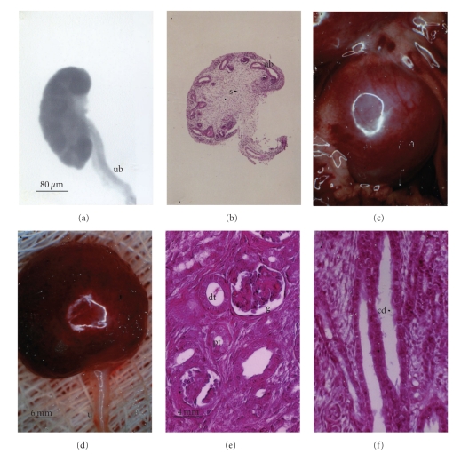
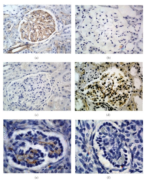
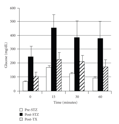
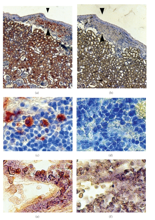
Lack of donor availability limits the number of human donor organs. The need for host immunosuppression complicates transplantation procedures. Ultrastructurally precise kidneys differentiate in situ following xenotransplantation in mesentery of embryonic pig renal primordia. The developing organ attracts its blood supply from the host, obviating humoral rejection. Engraftment of pig renal primordia transplanted directly into rats requires host immune suppression. However, insulin-producing cells originating from embryonic pig pancreas obtained very early following initiation of organogenesis [embryonic day 28 (E28)] engraft long term in nonimmune-suppressed diabetic rats or rhesus macaques. Engraftment of morphologically similar cells originating from adult porcine islets of Langerhans (islets) occurs in rats previously transplanted with E28 pig pancreatic primordia. Here, we review recent findings germane to xenotransplantation of pig renal or pancreatic primordia as a novel organ replacement strategy.


Figures

Figure 1
Figure 1
Photographs (a, c, d) and photomicrographs (b, e, f) of E28 pig renal primordia (a, b) or E28 pig renal primordia 7 weeks after transplantation into the mesentery of a rat (c–f). (a) E28 primordium (ub, ureteric bud); (b) E28 primordium (s, stroma; ub, ureteric bud); (c) E28 pig renal primordium 7 weeks after transplantation in a rat mesentery; (d) E28 pig renal primordium after removal from the mesentery (u, ureter), (e) Cortex with a glomerulus, (g) proximal tubule (pt), and distal tubule (dt) labeled; (f) Medulla with collecting duct (cd) labeled. Magnifications are shown for (a) and (b) (in (a)); (c) and (d) (in (d)); (e) and (f) (in (e)) reproduced with permission [8].
Figure 2
Figure 2
Photomicrographs of stained sections of (a, c) rat kidney; (b, d) pig kidney; (e, f) a pig renal primordium from an E28 embryo 8 weeks after transplantation into a rat mesentery, stained with RECA-1 (a, b, e) or CD31 (c, d, f). Glomerular capillaries in transplants stain positive for RECA-1 that is specific for rat endothelium (e) and negative for CD31 that is specific for pig endothelium (f). Magnifications are shown for (a–d) (in (a)) and (e) and (f) (in (e)), reproduced with permission [9].
Figure 3
Figure 3
Photomicrographs of mesenteric lymph node from an STZ-diabetic rhesus macaque, 78 days after transplantation of E28 pig pancreatic primordia. Sections (a), (c), and (e) are stained with an anti-insulin antibody. Sections (b), (e), and (f) are stained using a control serum. Sections of medullary sinus are delineated by arrows (a–d). Individual cells with beta cell morphology are delineated by arrowheads (e) and (f). Scale bars 120 um (a) and (b); 80 um (c) and (d); 20 um (e) and (f), reproduced with permission [11].
Figure 4
Figure 4
Intravenous glucose tolerance in rhesus macaques. Glucose in peripheral venous blood was measured prior to intravenous infusion of dextrose (Time 0) and at several times after infusion in three fasted rhesus macaques either prior to administration of STZ (Pre-STZ), 5 days following administration of STZ (Post-STZ), or 3 months following transplantation of 20–40 E28 pig pancreatic primordia in mesentery of STZ-diabetic macaques (Post-TX). Data are shown as mean ± SE (3 macaques), reproduced with permission [13].
Figure 5
Figure 5
Photomicrographs of kidney from a diabetic rat into which embryonic pig pancreas had been transplanted in mesentery, and pig islets had been transplanted subsequently in kidney stained using anti-insulin antibody (a, c) or control antibody (b, d) and sections hybridized to antisense (e) or sense (f) porcine proinsulin mRNA probes. Arrowheads delineate an expanded subcapsular space (a, b). Arrows delineate tissue in the subcapsular space that stains positive for insulin (red-brown) (c) or positive staining for porcine proinsulin mRNA (e). PT, proximal tubule (a, b). Scale bars 80 um (a, b) and 10 um (c–f), reproduced with permission from the American Society for Investigative Pathology [12].

Similar articles

Cited by

References

    1. Hammerman MR. Organogenesis of kidney and endocrine pancreas: the window opens. Organogenesis. 2007;3(2):59–66. - PMC - PubMed
    1. Hammerman MR. Xenotransplantation of pancreatic and kidney primordia: where do we stand? Transplant Immunology. 2009;21(2):93–100. - PMC - PubMed
    1. Brands K, Colvin E, Williams LJ, Wang R, Lock RB, Tuch BE. Reduced immunogenicity of first-trimester human fetal pancreas. Diabetes. 2008;57(3):627–634. - PubMed
    1. Dekel B, Amariglio N, Kaminski N, et al. Engraftment and differentiation of human metanephroi into functional mature nephrons after transplantation into mice is accompanied by a profile of gene expression similar to normal human kidney development. Journal of the American Society of Nephrology. 2002;13(4):977–990. - PubMed
    1. Dekel B, Burakova T, Arditti FD, et al. Human and porcine early kidney precursors as a new source for transplantation. Nature Medicine. 2003;9(1):53–60. - PubMed

LinkOut - more resources